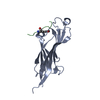
 Movie
Movie Controller


+ Open data

- Basic information
| Entry | Database: PDB / ID: 4xo1 | ||||||
|---|---|---|---|---|---|---|---|
| Title | crystal structure of Se-Met GnsA with double mutations | ||||||
 Components Components | Protein GnsA | ||||||
 Keywords Keywords | PROTEIN BINDING / suppressor | ||||||
| Function / homology | regulation of unsaturated fatty acid biosynthetic process / Gns / GnsA/GnsB toxin of bacterial toxin-antitoxin system / phosphatidylethanolamine biosynthetic process / protein homodimerization activity / identical protein binding / cytosol / Protein GnsA Function and homology information Function and homology information | ||||||
| Biological species |  | ||||||
| Method |  X-RAY DIFFRACTION / SYNCHROTRON / Resolution: 1.802 Å X-RAY DIFFRACTION / SYNCHROTRON / Resolution: 1.802 Å | ||||||
 Authors Authors | Zhan, L. / Gao, Z. / Dong, Y. | ||||||
 Citation Citation | Journal: Biochem.Biophys.Res.Commun. / Year: 2015 Title: Crystal structure of GnsA from Escherichia coli Authors: Wei, Y. / Zhan, L. / Gao, Z. / Prive, G.G. / Dong, Y. | ||||||
| History |
|
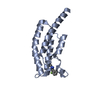
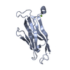
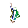
- Structure visualization
| Structure viewer | Molecule: MolmilJmol/JSmol |
|---|
- Downloads & links
-Download
| PDBx/mmCIF format | 4xo1.cif.gz | 26.6 KB | Display | PDBx/mmCIF format |
|---|---|---|---|---|
| PDB format | pdb4xo1.ent.gz | 15.8 KB | Display | PDB format |
| PDBx/mmJSON format | 4xo1.json.gz | Tree view | PDBx/mmJSON format | |
| Others |  Other downloads Other downloads |
-Validation report
| Arichive directory | https://data.pdbj.org/pub/pdb/validation_reports/xo/4xo1ftp://data.pdbj.org/pub/pdb/validation_reports/xo/4xo1 | HTTPS FTP |
|---|
-Related structure data


-Links
 PDBj
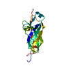
PDBj- Assembly
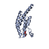
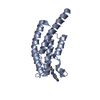
| Deposited unit | 
| ||||||||
|---|---|---|---|---|---|---|---|---|---|
| 1 | 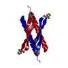
| ||||||||
| Unit cell |
|
-Components
| #1: Protein | Mass: 7236.585 Da / Num. of mol.: 1 / Mutation: L25M, I36M Source method: isolated from a genetically manipulated source Source: (gene. exp.) |
|---|---|
| #2: Water | ChemComp-HOH /  Mass: 18.015 Da / Num. of mol.: 44 / Source method: isolated from a natural source / Formula: H2O Mass: 18.015 Da / Num. of mol.: 44 / Source method: isolated from a natural source / Formula: H2O |
| Has protein modification | Y |
-Experimental details
-Experiment
| Experiment | Method: X-RAY DIFFRACTION |
|---|
- Sample preparation
| Crystal | Density Matthews: 2.25 Å3/Da / Density % sol: 45.4 % Description: THE ENTRY CONTAINS FRIEDEL PAIRS IN F_PLUS/MINUS COLUMNS. |
|---|---|
| Crystal grow | Temperature: 293 K / Method: vapor diffusion, sitting drop / pH: 5.5 Details: 0.1M BIS-TRIS pH 5.5, 25%(w/v) Polyethylene glycol 3,350 |
-Data collection
| Diffraction | Mean temperature: 100 K |
|---|---|
| Diffraction source | Source: SYNCHROTRON / Site: BSRF  / Beamline: 3W1A / Wavelength: 0.9792 Å / Beamline: 3W1A / Wavelength: 0.9792 Å |
| Detector | Type: MAR CCD 165 mm / Detector: CCD / Date: Oct 5, 2014 |
| Radiation | Protocol: SINGLE WAVELENGTH / Monochromatic (M) / Laue (L): M / Scattering type: x-ray |
| Radiation wavelength | Wavelength: 0.9792 Å / Relative weight: 1 |
| Reflection | Resolution: 1.8→50 Å / Num. obs: 10526 / % possible obs: 99.9 % / Redundancy: 14.1 % / Rmerge(I) obs: 0.06 / Net I/σ(I): 60.3 |
| Reflection shell | Resolution: 1.8→1.83 Å / Redundancy: 14.2 % / Rmerge(I) obs: 0.414 / Mean I/σ(I) obs: 6.9 / % possible all: 100 |
- Processing
| Software |
| ||||||||||||||||||||||||||||
|---|---|---|---|---|---|---|---|---|---|---|---|---|---|---|---|---|---|---|---|---|---|---|---|---|---|---|---|---|---|
| Refinement | Resolution: 1.802→28.37 Å / SU ML: 0.25 / Cross valid method: FREE R-VALUE / σ(F): 1.34 / Phase error: 30.01 / Stereochemistry target values: ML Details: SF FILE CONTAINS FRIEDEL PAIRS UNDER I/F_MINUS AND I/F_PLUS COLUMNS.
| ||||||||||||||||||||||||||||
| Solvent computation | Shrinkage radii: 0.9 Å / VDW probe radii: 1.11 Å / Solvent model: FLAT BULK SOLVENT MODEL | ||||||||||||||||||||||||||||
| Refinement step | Cycle: LAST / Resolution: 1.802→28.37 Å
| ||||||||||||||||||||||||||||
| Refine LS restraints |
| ||||||||||||||||||||||||||||
| LS refinement shell |
|
