 Movie
Movie Controller

[English] 日本語
 Yorodumi
- PDB-4xeg: Structure of the enzyme-product complex resulting from TDG action... -
+ Open data

- Basic information
| Entry | Database: PDB / ID: 4xeg | ||||||
|---|---|---|---|---|---|---|---|
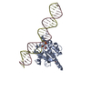
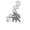
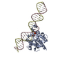
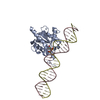
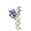
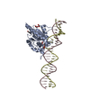
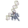
| Title | Structure of the enzyme-product complex resulting from TDG action on a G/hmU mismatch | ||||||
 Components Components |
| ||||||
 Keywords Keywords | HYDROLASE/DNA / DNA glycosylase / abasic site / enzyme-product complex / 5-hydroxymethyluracil / HYDROLASE-DNA complex | ||||||
| Function / homology |  Function and homology information Function and homology informationG/U mismatch-specific uracil-DNA glycosylase activity / thymine-DNA glycosylase / G/T mismatch-specific thymine-DNA glycosylase activity / chromosomal 5-methylcytosine DNA demethylation, oxidation pathway / TET1,2,3 and TDG demethylate DNA / pyrimidine-specific mismatch base pair DNA N-glycosylase activity / base-excision repair, AP site formation / depyrimidination / DNA N-glycosylase activity / sodium ion binding ...G/U mismatch-specific uracil-DNA glycosylase activity / thymine-DNA glycosylase / G/T mismatch-specific thymine-DNA glycosylase activity / chromosomal 5-methylcytosine DNA demethylation, oxidation pathway / TET1,2,3 and TDG demethylate DNA / pyrimidine-specific mismatch base pair DNA N-glycosylase activity / base-excision repair, AP site formation / depyrimidination / DNA N-glycosylase activity / sodium ion binding / mismatched DNA binding / Displacement of DNA glycosylase by APEX1 / SUMO binding / uracil DNA N-glycosylase activity / chloride ion binding / regulation of embryonic development / SUMOylation of DNA damage response and repair proteins / epigenetic regulation of gene expression / Recognition and association of DNA glycosylase with site containing an affected pyrimidine / Cleavage of the damaged pyrimidine / protein kinase C binding / transcription coregulator activity / PML body / base-excision repair / double-stranded DNA binding / DNA-binding transcription factor binding / nucleic acid binding / damaged DNA binding / protein domain specific binding / magnesium ion binding / negative regulation of transcription by RNA polymerase II / DNA binding / nucleoplasm / ATP binding / nucleus / plasma membrane Similarity search - Function | ||||||
| Biological species |  Homo sapiens (human) Homo sapiens (human)unidentified (others) | ||||||
| Method |  X-RAY DIFFRACTION / SYNCHROTRON / MOLECULAR REPLACEMENT / molecular replacement / Resolution: 1.72 Å X-RAY DIFFRACTION / SYNCHROTRON / MOLECULAR REPLACEMENT / molecular replacement / Resolution: 1.72 Å | ||||||
 Authors Authors | Pozharski, E. / Malik, S.S. / Drohat, A.C. | ||||||
| Funding support |  United States, 1items United States, 1items
| ||||||
 Citation Citation | Journal: Nucleic Acids Res. / Year: 2015 Title: Thymine DNA glycosylase exhibits negligible affinity for nucleobases that it removes from DNA. Authors: Malik, S.S. / Coey, C.T. / Varney, K.M. / Pozharski, E. / Drohat, A.C. | ||||||
| History |
|
- Structure visualization
| Structure viewer | Molecule: MolmilJmol/JSmol |
|---|
- Downloads & links
-Download
| PDBx/mmCIF format | 4xeg.cif.gz | 98.2 KB | Display | PDBx/mmCIF format |
|---|---|---|---|---|
| PDB format | pdb4xeg.ent.gz | 68.5 KB | Display | PDB format |
| PDBx/mmJSON format | 4xeg.json.gz | Tree view | PDBx/mmJSON format | |
| Others |  Other downloads Other downloads |
-Validation report
| Arichive directory | https://data.pdbj.org/pub/pdb/validation_reports/xe/4xegftp://data.pdbj.org/pub/pdb/validation_reports/xe/4xeg | HTTPS FTP |
|---|
-Related structure data
| Related structure data |  4z3aC  4z47C  4z7bC  4z7zC 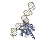 4fncS S: Starting model for refinement C: citing same article ( |
|---|---|
| Similar structure data |


-Links
 PDBj
PDBj


- Assembly
| Deposited unit | 
| ||||||||
|---|---|---|---|---|---|---|---|---|---|
| 1 |
| ||||||||
| Unit cell |
| ||||||||
| Components on special symmetry positions |
|
-Components
-Protein , 1 types, 1 molecules A
| #1: Protein | Mass: 23070.670 Da / Num. of mol.: 1 Source method: isolated from a genetically manipulated source Source: (gene. exp.) Homo sapiens (human) / Gene: TDG / Production host:  |
|---|
-DNA chain , 2 types, 2 molecules CD
| #2: DNA chain | Mass: 8646.565 Da / Num. of mol.: 1 / Source method: obtained synthetically / Source: (synth.) unidentified (others) |
|---|---|
| #3: DNA chain | Mass: 8473.431 Da / Num. of mol.: 1 / Source method: obtained synthetically / Source: (synth.) unidentified (others) |
-Non-polymers , 3 types, 305 molecules 


| #4: Chemical |  Mass: 62.068 Da / Num. of mol.: 2 / Source method: obtained synthetically / Formula: C2H6O2 Mass: 62.068 Da / Num. of mol.: 2 / Source method: obtained synthetically / Formula: C2H6O2#5: Chemical |  Mass: 60.052 Da / Num. of mol.: 3 / Source method: obtained synthetically / Formula: C2H4O2 Mass: 60.052 Da / Num. of mol.: 3 / Source method: obtained synthetically / Formula: C2H4O2#6: Water | ChemComp-HOH / | Mass: 18.015 Da / Num. of mol.: 300 / Source method: isolated from a natural source / Formula: H2O |
|---|
-Experimental details
-Experiment
| Experiment | Method: X-RAY DIFFRACTION / Number of used crystals: 1 |
|---|
- Sample preparation
| Crystal | Density Matthews: 2.47 Å3/Da / Density % sol: 50.27 % |
|---|---|
| Crystal grow | Temperature: 295 K / Method: vapor diffusion, sitting drop / pH: 6 Details: 20% (w/v) PEG 4000, 0.2 M ammonium acetate, 0.1 M sodium acetate, pH 6.0 |
-Data collection
| Diffraction | Mean temperature: 100 K | |||||||||||||||||||||||||||
|---|---|---|---|---|---|---|---|---|---|---|---|---|---|---|---|---|---|---|---|---|---|---|---|---|---|---|---|---|
| Diffraction source | Source: SYNCHROTRON / Site: SSRL / Beamline: BL7-1 / Wavelength: 0.97948 Å | |||||||||||||||||||||||||||
| Detector | Type: ADSC QUANTUM 315r / Detector: CCD / Date: Jun 10, 2014 | |||||||||||||||||||||||||||
| Radiation | Protocol: SINGLE WAVELENGTH / Monochromatic (M) / Laue (L): M / Scattering type: x-ray | |||||||||||||||||||||||||||
| Radiation wavelength | Wavelength: 0.97948 Å / Relative weight: 1 | |||||||||||||||||||||||||||
| Reflection | Resolution: 1.72→39.14 Å / Num. obs: 40036 / % possible obs: 97.2 % / Redundancy: 6.6 % / Biso Wilson estimate: 29.1 Å2 / CC1/2: 1 / Rmerge(I) obs: 0.038 / Rpim(I) all: 0.016 / Net I/σ(I): 23.8 / Num. measured all: 266180 | |||||||||||||||||||||||||||
| Reflection shell | Diffraction-ID: 1 / Rejects: _
|
-Phasing
| Phasing | Method: molecular replacement |
|---|
- Processing
| Software |
| |||||||||||||||||||||||||||||||||||||||||||||||||||||||||||||||||||||||||||
|---|---|---|---|---|---|---|---|---|---|---|---|---|---|---|---|---|---|---|---|---|---|---|---|---|---|---|---|---|---|---|---|---|---|---|---|---|---|---|---|---|---|---|---|---|---|---|---|---|---|---|---|---|---|---|---|---|---|---|---|---|---|---|---|---|---|---|---|---|---|---|---|---|---|---|---|---|
| Refinement | Method to determine structure: MOLECULAR REPLACEMENT Starting model: PDB ENTRY 4FNC Resolution: 1.72→39.14 Å / Cor.coef. Fo:Fc: 0.969 / Cor.coef. Fo:Fc free: 0.953 / SU B: 3.258 / SU ML: 0.099 / Cross valid method: THROUGHOUT / σ(F): 0 / ESU R: 0.115 / ESU R Free: 0.117 / Stereochemistry target values: MAXIMUM LIKELIHOOD Details: HYDROGENS HAVE BEEN ADDED IN THE RIDING POSITIONS U VALUES : REFINED INDIVIDUALLY
| |||||||||||||||||||||||||||||||||||||||||||||||||||||||||||||||||||||||||||
| Solvent computation | Ion probe radii: 0.8 Å / Shrinkage radii: 0.8 Å / VDW probe radii: 1.2 Å / Solvent model: MASK | |||||||||||||||||||||||||||||||||||||||||||||||||||||||||||||||||||||||||||
| Displacement parameters | Biso max: 100.21 Å2 / Biso mean: 38.544 Å2 / Biso min: 17.31 Å2
| |||||||||||||||||||||||||||||||||||||||||||||||||||||||||||||||||||||||||||
| Refinement step | Cycle: final / Resolution: 1.72→39.14 Å
| |||||||||||||||||||||||||||||||||||||||||||||||||||||||||||||||||||||||||||
| Refine LS restraints |
| |||||||||||||||||||||||||||||||||||||||||||||||||||||||||||||||||||||||||||
| LS refinement shell | Resolution: 1.718→1.763 Å / Total num. of bins used: 20
|
