Monochromator: SI(111) / Protocol: SINGLE WAVELENGTH / Monochromatic (M) / Laue (L): M / Scattering type: x-ray
Radiation wavelength
Wavelength: 0.97924 Å / Relative weight: 1
Reflection
Resolution: 1.23→20 Å / Num. obs: 25859 / % possible obs: 98.4 % / Observed criterion σ(I): -3 / Redundancy: 3.6 % / Rmerge(I) obs: 0.116 / Net I/σ(I): 12.81
Reflection shell
Resolution: 1.23→1.25 Å / Redundancy: 2.7 % / Rmerge(I) obs: 0.48 / Mean I/σ(I) obs: 1.98 / % possible all: 89.4
-
Processing
Software
Name
Version
Classification
PHENIX
(phenix.refine: 1.9_1692)
refinement
HKL-3000
phasing
HKL-3000
datascaling
SBC-Collect
datacollection
SHELX
phasing
MLPHARE
phasing
DM
phasing
ARP
modelbuilding
HKL-3000
datareduction
Refinement
Method to determine structure: SAD / Resolution: 1.23→19.9 Å / SU ML: 0.11 / Cross valid method: THROUGHOUT / σ(F): 0 / Phase error: 16.48 / Stereochemistry target values: ML Details: HYDROGEN ATOMS HAVE BEEN ADDED IN RIDING POSITIONS, UNMODELED DENSITY NEAR THR89 POSSIBLY CORRESPONDS TO CALCIUM ION BUT DUE TO INCOMPLETE AND INCOSISTENT COORDINATION IT COULD NOT BE MODELED WITH CONFIDENCE.
Rfactor
Num. reflection
% reflection
Selection details
Rfree
0.1687
1058
4.09 %
random
Rwork
0.1382
-
-
-
obs
0.1394
25851
98.09 %
-
Solvent computation
Shrinkage radii: 0.9 Å / VDW probe radii: 1.11 Å / Solvent model: FLAT BULK SOLVENT MODEL
Refinement step
Cycle: LAST / Resolution: 1.23→19.9 Å
Protein
Nucleic acid
Ligand
Solvent
Total
Num. atoms
769
0
9
123
901
Refine LS restraints
Refine-ID
Type
Dev ideal
Number
X-RAY DIFFRACTION
f_bond_d
0.012
863
X-RAY DIFFRACTION
f_angle_d
1.435
1187
X-RAY DIFFRACTION
f_dihedral_angle_d
12.94
307
X-RAY DIFFRACTION
f_chiral_restr
0.087
124
X-RAY DIFFRACTION
f_plane_restr
0.009
157
LS refinement shell
Resolution (Å)
Rfactor Rfree
Num. reflection Rfree
Rfactor Rwork
Num. reflection Rwork
Refine-ID
% reflection obs (%)
1.23-1.2833
0.195
112
0.1747
2803
X-RAY DIFFRACTION
90
1.2833-1.351
0.2587
143
0.1763
3026
X-RAY DIFFRACTION
98
1.351-1.4356
0.2044
139
0.1583
3107
X-RAY DIFFRACTION
99
1.4356-1.5464
0.1949
120
0.1402
3119
X-RAY DIFFRACTION
100
1.5464-1.7019
0.1653
133
0.1302
3127
X-RAY DIFFRACTION
100
1.7019-1.948
0.1495
136
0.1148
3152
X-RAY DIFFRACTION
100
1.948-2.4536
0.149
135
0.1208
3199
X-RAY DIFFRACTION
100
2.4536-19.9068
0.1583
140
0.1441
3260
X-RAY DIFFRACTION
99
+
About Yorodumi
-
News
-
Feb 9, 2022. New format data for meta-information of EMDB entries
New format data for meta-information of EMDB entries
Version 3 of the EMDB header file is now the official format.
The previous official version 1.9 will be removed from the archive.
In the structure databanks used in Yorodumi, some data are registered as the other names, "COVID-19 virus" and "2019-nCoV". Here are the details of the virus and the list of structure data.
Jan 31, 2019. EMDB accession codes are about to change! (news from PDBe EMDB page)
EMDB accession codes are about to change! (news from PDBe EMDB page)
The allocation of 4 digits for EMDB accession codes will soon come to an end. Whilst these codes will remain in use, new EMDB accession codes will include an additional digit and will expand incrementally as the available range of codes is exhausted. The current 4-digit format prefixed with “EMD-” (i.e. EMD-XXXX) will advance to a 5-digit format (i.e. EMD-XXXXX), and so on. It is currently estimated that the 4-digit codes will be depleted around Spring 2019, at which point the 5-digit format will come into force.
The EM Navigator/Yorodumi systems omit the EMD- prefix.
Related info.:Q: What is EMD? / ID/Accession-code notation in Yorodumi/EM Navigator
Yorodumi is a browser for structure data from EMDB, PDB, SASBDB, etc.
This page is also the successor to EM Navigator detail page, and also detail information page/front-end page for Omokage search.
The word "yorodu" (or yorozu) is an old Japanese word meaning "ten thousand". "mi" (miru) is to see.
Related info.:EMDB / PDB / SASBDB / Comparison of 3 databanks / Yorodumi Search / Aug 31, 2016. New EM Navigator & Yorodumi / Yorodumi Papers / Jmol/JSmol / Function and homology information / Changes in new EM Navigator and Yorodumi
 Movie
Movie Controller

 Yorodumi
 Open data
 Basic information
 Components
 Keywords
 Function and homology information
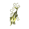
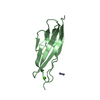
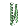
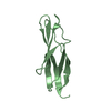
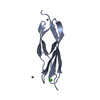
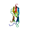
 Thermoplasmatales archaeon SCGC AB-540-F20 (archaea)
 X-RAY DIFFRACTION /
  Authors
 United States, 1items
  Citation
 Structure visualization
 Downloads & links
 Other downloads
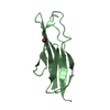


 PDBj
PDBj
 Assembly
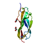


 Mass: 40.078 Da / Num. of mol.: 1 / Source method: obtained synthetically / Formula: Ca
 Mass: 62.068 Da / Num. of mol.: 2 / Source method: obtained synthetically / Formula: C2H6O2
 Mass: 18.015 Da / Num. of mol.: 123 / Source method: isolated from a natural source / Formula: H2O
 Sample preparation
 Processing