 Movie
Movie Controller


+ Open data

- Basic information
| Entry | Database: PDB / ID: 4x0x | ||||||
|---|---|---|---|---|---|---|---|
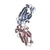
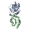
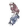
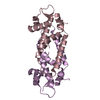
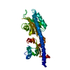
| Title | The structure of AhpE from Mycobacterium tuberculosis revisited | ||||||
 Components Components | Putative peroxiredoxin MT2298 | ||||||
 Keywords Keywords | OXIDOREDUCTASE / Peroxiredoxins / Protein Structure | ||||||
| Function / homology |  Function and homology information Function and homology information | ||||||
| Biological species |   Mycobacterium tuberculosis (bacteria) Mycobacterium tuberculosis (bacteria) | ||||||
| Method |  X-RAY DIFFRACTION / SYNCHROTRON / MOLECULAR REPLACEMENT / Resolution: 1.9 Å X-RAY DIFFRACTION / SYNCHROTRON / MOLECULAR REPLACEMENT / Resolution: 1.9 Å | ||||||
 Authors Authors | Pallo, A. / Messens, J. | ||||||
| Funding support |  Belgium, 1items Belgium, 1items
| ||||||
 Citation Citation | Journal: Sci Rep / Year: 2016 Title: Revisiting sulfur H-bonds in proteins: The example of peroxiredoxin AhpE. Authors: van Bergen, L.A. / Alonso, M. / Pallo, A. / Nilsson, L. / De Proft, F. / Messens, J. #1: Journal: J. Mol. Biol. / Year: 2005Title: Crystal Structure of AhpE from Mycobacterium tuberculosis, a 1-Cys peroxiredoxin. Authors: Li, S. / Peterson, N.A. / Kim, M.Y. / Kim, C.Y. / Hung, L.W. / Yu, M. / Lekin, T. / Segelke, B.W. / Lott, J.S. / Baker, E.N. | ||||||
| History |
|
- Structure visualization
| Structure viewer | Molecule: MolmilJmol/JSmol |
|---|
- Downloads & links
-Download
| PDBx/mmCIF format | 4x0x.cif.gz | 240.9 KB | Display | PDBx/mmCIF format |
|---|---|---|---|---|
| PDB format | pdb4x0x.ent.gz | 195.1 KB | Display | PDB format |
| PDBx/mmJSON format | 4x0x.json.gz | Tree view | PDBx/mmJSON format | |
| Others |  Other downloads Other downloads |
-Validation report
| Arichive directory | https://data.pdbj.org/pub/pdb/validation_reports/x0/4x0xftp://data.pdbj.org/pub/pdb/validation_reports/x0/4x0x | HTTPS FTP |
|---|
-Related structure data
| Related structure data | 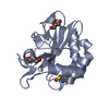 4x1uC  1xvwS S: Starting model for refinement C: citing same article ( |
|---|---|
| Similar structure data |


-Links
 PDBj
PDBj- Assembly
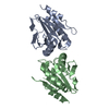
| Deposited unit | 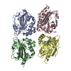
| ||||||||||||||||||||||||||||||||||||||||||||||||||||||||||||||||||||||||||||||||||||||||||||||||||||||||||||||||||||||||||||||||||||||||||||||||||||||
|---|---|---|---|---|---|---|---|---|---|---|---|---|---|---|---|---|---|---|---|---|---|---|---|---|---|---|---|---|---|---|---|---|---|---|---|---|---|---|---|---|---|---|---|---|---|---|---|---|---|---|---|---|---|---|---|---|---|---|---|---|---|---|---|---|---|---|---|---|---|---|---|---|---|---|---|---|---|---|---|---|---|---|---|---|---|---|---|---|---|---|---|---|---|---|---|---|---|---|---|---|---|---|---|---|---|---|---|---|---|---|---|---|---|---|---|---|---|---|---|---|---|---|---|---|---|---|---|---|---|---|---|---|---|---|---|---|---|---|---|---|---|---|---|---|---|---|---|---|---|---|---|
| 1 | 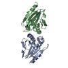
| ||||||||||||||||||||||||||||||||||||||||||||||||||||||||||||||||||||||||||||||||||||||||||||||||||||||||||||||||||||||||||||||||||||||||||||||||||||||
| 2 | 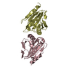
| ||||||||||||||||||||||||||||||||||||||||||||||||||||||||||||||||||||||||||||||||||||||||||||||||||||||||||||||||||||||||||||||||||||||||||||||||||||||
| Unit cell |
| ||||||||||||||||||||||||||||||||||||||||||||||||||||||||||||||||||||||||||||||||||||||||||||||||||||||||||||||||||||||||||||||||||||||||||||||||||||||
| Noncrystallographic symmetry (NCS) | NCS domain:
NCS domain segments: Component-ID: _ / Beg auth comp-ID: HIS / Beg label comp-ID: HIS / Refine code: _
NCS ensembles :
|
-Components
| #1: Protein | Mass: 16972.098 Da / Num. of mol.: 4 Source method: isolated from a genetically manipulated source Source: (gene. exp.) Mycobacterium tuberculosis (bacteria) / Strain: CDC 1551 / Oshkosh / Gene: MT2298 / Production host: #2: Water | ChemComp-HOH / |  Mass: 18.015 Da / Num. of mol.: 311 / Source method: isolated from a natural source / Formula: H2O Mass: 18.015 Da / Num. of mol.: 311 / Source method: isolated from a natural source / Formula: H2O |
|---|
-Experimental details
-Experiment
| Experiment | Method: X-RAY DIFFRACTION / Number of used crystals: 1 |
|---|
- Sample preparation
| Crystal | Density Matthews: 2.74 Å3/Da / Density % sol: 55.09 % |
|---|---|
| Crystal grow | Temperature: 291 K / Method: vapor diffusion, sitting drop / pH: 4.5 Details: 1.8M sodium malonate pH 5.0 mixed with 0.1M sodium acetate pH 4.5, VAPOR DIFFUSION, SITTING DROP, temperature 291K |
-Data collection
| Diffraction | Mean temperature: 113 K |
|---|---|
| Diffraction source | Source: SYNCHROTRON / Site: ALS  / Beamline: 5.0.2 / Wavelength: 0.91983 Å / Beamline: 5.0.2 / Wavelength: 0.91983 Å |
| Detector | Type: ADSC QUANTUM 210 / Detector: CCD |
| Radiation | Protocol: SINGLE WAVELENGTH / Monochromatic (M) / Laue (L): M / Scattering type: x-ray |
| Radiation wavelength | Wavelength: 0.91983 Å / Relative weight: 1 |
| Reflection | Resolution: 1.9→40 Å / Num. obs: 56084 / % possible obs: 99.83 % / Rsym value: 0.082 / Net I/σ(I): 16.2 |
| Reflection shell | Resolution: 1.9→1.97 Å / Mean I/σ(I) obs: 2.29 / Num. unique all: 5817 / Rsym value: 0.598 / % possible all: 99.6 |
- Processing
| Software |
| |||||||||||||||||||||||||||||||||||||||||||||||||||||||||||||||||||||||||||
|---|---|---|---|---|---|---|---|---|---|---|---|---|---|---|---|---|---|---|---|---|---|---|---|---|---|---|---|---|---|---|---|---|---|---|---|---|---|---|---|---|---|---|---|---|---|---|---|---|---|---|---|---|---|---|---|---|---|---|---|---|---|---|---|---|---|---|---|---|---|---|---|---|---|---|---|---|
| Refinement | Method to determine structure: MOLECULAR REPLACEMENT Starting model: 1xvw Resolution: 1.9→40 Å / Cor.coef. Fo:Fc: 0.914 / Cor.coef. Fo:Fc free: 0.891 / WRfactor Rfree: 0.2358 / WRfactor Rwork: 0.2073 / FOM work R set: 0.8068 / SU B: 7.824 / SU ML: 0.115 / SU R Cruickshank DPI: 0.16 / SU Rfree: 0.1453 / Cross valid method: THROUGHOUT / σ(F): 0 / ESU R: 0.16 / ESU R Free: 0.145 / Stereochemistry target values: MAXIMUM LIKELIHOOD / Details: HYDROGENS HAVE BEEN ADDED IN THE RIDING POSITIONS
| |||||||||||||||||||||||||||||||||||||||||||||||||||||||||||||||||||||||||||
| Solvent computation | Ion probe radii: 0.8 Å / Shrinkage radii: 0.8 Å / VDW probe radii: 1.4 Å / Solvent model: BABINET MODEL WITH MASK | |||||||||||||||||||||||||||||||||||||||||||||||||||||||||||||||||||||||||||
| Displacement parameters | Biso max: 84.94 Å2 / Biso mean: 23.74 Å2 / Biso min: 11.78 Å2
| |||||||||||||||||||||||||||||||||||||||||||||||||||||||||||||||||||||||||||
| Refinement step | Cycle: final / Resolution: 1.9→40 Å
| |||||||||||||||||||||||||||||||||||||||||||||||||||||||||||||||||||||||||||
| Refine LS restraints |
| |||||||||||||||||||||||||||||||||||||||||||||||||||||||||||||||||||||||||||
| Refine LS restraints NCS | Refine-ID: X-RAY DIFFRACTION / Type: interatomic distance / Weight position: 0.05
| |||||||||||||||||||||||||||||||||||||||||||||||||||||||||||||||||||||||||||
| LS refinement shell | Resolution: 1.9→1.949 Å / Total num. of bins used: 20
| |||||||||||||||||||||||||||||||||||||||||||||||||||||||||||||||||||||||||||
| Refinement TLS params. | Method: refined / Origin x: -36.17 Å / Origin y: 38.1751 Å / Origin z: 11.6834 Å
| |||||||||||||||||||||||||||||||||||||||||||||||||||||||||||||||||||||||||||
| Refinement TLS group |
|
