 Movie
Movie Controller

[English] 日本語
 Yorodumi
- PDB-4wux: Crystal Structure of Surfactant Protein-A DED Mutant (E171D/P175E... -
+ Open data

- Basic information
| Entry | Database: PDB / ID: 4wux | ||||||
|---|---|---|---|---|---|---|---|
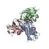
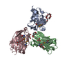
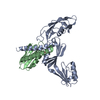
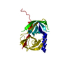
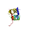
| Title | Crystal Structure of Surfactant Protein-A DED Mutant (E171D/P175E/K203D) Complexed with Mannose | ||||||
 Components Components | Pulmonary surfactant-associated protein A | ||||||
 Keywords Keywords | SUGAR BINDING PROTEIN / COLLECTIN / CARBOHYDRATE BINDING / LECTIN / LIPID BINDING | ||||||
| Function / homology |  Function and homology information Function and homology informationSignal regulatory protein family interactions / Surfactant metabolism / Toll Like Receptor 4 (TLR4) Cascade / Regulation of TLR by endogenous ligand / opsonization / response to interleukin-6 / respiratory gaseous exchange by respiratory system / collagen trimer / response to epidermal growth factor / response to vitamin A ...Signal regulatory protein family interactions / Surfactant metabolism / Toll Like Receptor 4 (TLR4) Cascade / Regulation of TLR by endogenous ligand / opsonization / response to interleukin-6 / respiratory gaseous exchange by respiratory system / collagen trimer / response to epidermal growth factor / response to vitamin A / response to hyperoxia / response to retinoic acid / multivesicular body / response to hormone / cellular response to nitric oxide / positive regulation of phagocytosis / response to glucocorticoid / cellular response to mechanical stimulus / circadian rhythm / carbohydrate binding / response to lipopolysaccharide / response to hypoxia / : / metal ion binding Similarity search - Function | ||||||
| Biological species |  | ||||||
| Method |  X-RAY DIFFRACTION / FOURIER SYNTHESIS / Resolution: 1.9 Å X-RAY DIFFRACTION / FOURIER SYNTHESIS / Resolution: 1.9 Å | ||||||
 Authors Authors | Rynkiewicz, M.J. / Wu, H. / Cafarella, T.R. / Nikolaidis, N.M. / Head, J.F. / Seaton, B.A. / McCormack, F.X. | ||||||
| Funding support |  United States, 1items United States, 1items
| ||||||
 Citation Citation | Journal: To be published Title: Differential ligand binding specificities of the pulmonary collectins are determined by the conformational freedom of a surface loop Authors: Rynkiewicz, M.J. / Wu, H. / Cafarella, T.R. / Nikolaidis, N.M. / Head, J.F. / Seaton, B.A. / McCormack, F.X. | ||||||
| History |
|
- Structure visualization
| Structure viewer | Molecule: MolmilJmol/JSmol |
|---|
- Downloads & links
-Download
| PDBx/mmCIF format | 4wux.cif.gz | 78.6 KB | Display | PDBx/mmCIF format |
|---|---|---|---|---|
| PDB format | pdb4wux.ent.gz | 57.6 KB | Display | PDB format |
| PDBx/mmJSON format | 4wux.json.gz | Tree view | PDBx/mmJSON format | |
| Others |  Other downloads Other downloads |
-Validation report
| Arichive directory | https://data.pdbj.org/pub/pdb/validation_reports/wu/4wuxftp://data.pdbj.org/pub/pdb/validation_reports/wu/4wux | HTTPS FTP |
|---|
-Related structure data
| Related structure data |  4wr9SC  4wrcC  4wreC  4wrfC  4wuwC C: citing same article ( S: Starting model for refinement |
|---|---|
| Similar structure data |

-Links
 PDBj
PDBj


- Assembly
| Deposited unit | 
| ||||||||||||
|---|---|---|---|---|---|---|---|---|---|---|---|---|---|
| 1 | 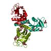
| ||||||||||||
| Unit cell |
| ||||||||||||
| Components on special symmetry positions |
|
-Components
| #1: Protein | Mass: 16724.461 Da / Num. of mol.: 1 / Fragment: neck and carbohydrate recognition domain / Mutation: E171D/P175E//N187S/K203D Source method: isolated from a genetically manipulated source Source: (gene. exp.)  Trichoplusia ni (cabbage looper) / References: UniProt: P08427 Trichoplusia ni (cabbage looper) / References: UniProt: P08427 | ||||||
|---|---|---|---|---|---|---|---|
| #2: Sugar |   Type: D-saccharide, alpha linking / Mass: 180.156 Da / Num. of mol.: 3 / Source method: obtained synthetically / Formula: C6H12O6 Type: D-saccharide, alpha linking / Mass: 180.156 Da / Num. of mol.: 3 / Source method: obtained synthetically / Formula: C6H12O6#3: Chemical |   Mass: 40.078 Da / Num. of mol.: 2 / Source method: obtained synthetically / Formula: Ca Mass: 40.078 Da / Num. of mol.: 2 / Source method: obtained synthetically / Formula: Ca#4: Water | ChemComp-HOH / |  Mass: 18.015 Da / Num. of mol.: 77 / Source method: isolated from a natural source / Formula: H2O Mass: 18.015 Da / Num. of mol.: 77 / Source method: isolated from a natural source / Formula: H2OHas protein modification | Y | |
-Experimental details
-Experiment
| Experiment | Method: X-RAY DIFFRACTION / Number of used crystals: 1 |
|---|
- Sample preparation
| Crystal | Density Matthews: 3.65 Å3/Da / Density % sol: 66.3 % |
|---|---|
| Crystal grow | Temperature: 290 K / Method: vapor diffusion, hanging drop / pH: 6 Details: The crystal was grown in hanging drops using 10 mM sodium cacodylate (pH 6.0), 80 mM calcium acetate, 0-10% glycerol, and 4-10% (w/v) PEG 20,000 as a reservoir. Before freezing for x-ray ...Details: The crystal was grown in hanging drops using 10 mM sodium cacodylate (pH 6.0), 80 mM calcium acetate, 0-10% glycerol, and 4-10% (w/v) PEG 20,000 as a reservoir. Before freezing for x-ray data collection, crystal was soaked in reservoir solution of 10 mM calcium acetate without glycerol, but with stepwise additions of 5, 10, and 15% 2-methyl-2,4-pentanediol with 25% mannose for 10 minutes at each step |
-Data collection
| Diffraction | Mean temperature: 100 K | ||||||||||||||||||||||||||||||||||||||||||||||||||||||||||||||||||
|---|---|---|---|---|---|---|---|---|---|---|---|---|---|---|---|---|---|---|---|---|---|---|---|---|---|---|---|---|---|---|---|---|---|---|---|---|---|---|---|---|---|---|---|---|---|---|---|---|---|---|---|---|---|---|---|---|---|---|---|---|---|---|---|---|---|---|---|
| Diffraction source | Source: ROTATING ANODE / Type: RIGAKU RU300 / Wavelength: 1.5418 Å | ||||||||||||||||||||||||||||||||||||||||||||||||||||||||||||||||||
| Detector | Type: RIGAKU RAXIS IV / Detector: IMAGE PLATE / Date: Mar 21, 2012 | ||||||||||||||||||||||||||||||||||||||||||||||||||||||||||||||||||
| Radiation | Protocol: SINGLE WAVELENGTH / Monochromatic (M) / Laue (L): M / Scattering type: x-ray | ||||||||||||||||||||||||||||||||||||||||||||||||||||||||||||||||||
| Radiation wavelength | Wavelength: 1.5418 Å / Relative weight: 1 | ||||||||||||||||||||||||||||||||||||||||||||||||||||||||||||||||||
| Reflection | Resolution: 1.9→15 Å / Num. obs: 18934 / % possible obs: 98.5 % / Redundancy: 5.2 % / Biso Wilson estimate: 35.47 Å2 / Rmerge(I) obs: 0.052 / Χ2: 1.257 / Net I/av σ(I): 30.366 / Net I/σ(I): 23.8 / Num. measured all: 98492 | ||||||||||||||||||||||||||||||||||||||||||||||||||||||||||||||||||
| Reflection shell | Diffraction-ID: 1 / Rejects: _
|
- Processing
| Software |
| |||||||||||||||||||||||||||||||||||||||||||||||||||||||||||||||||||||||||||||||||||||||||||||||||||||||||
|---|---|---|---|---|---|---|---|---|---|---|---|---|---|---|---|---|---|---|---|---|---|---|---|---|---|---|---|---|---|---|---|---|---|---|---|---|---|---|---|---|---|---|---|---|---|---|---|---|---|---|---|---|---|---|---|---|---|---|---|---|---|---|---|---|---|---|---|---|---|---|---|---|---|---|---|---|---|---|---|---|---|---|---|---|---|---|---|---|---|---|---|---|---|---|---|---|---|---|---|---|---|---|---|---|---|---|
| Refinement | Method to determine structure: FOURIER SYNTHESIS Starting model: PDB entry 4WR9 Resolution: 1.9→14.963 Å / FOM work R set: 0.8841 / SU ML: 0.19 / Cross valid method: FREE R-VALUE / σ(F): 1.35 / Phase error: 18.75 / Stereochemistry target values: ML
| |||||||||||||||||||||||||||||||||||||||||||||||||||||||||||||||||||||||||||||||||||||||||||||||||||||||||
| Solvent computation | Shrinkage radii: 0.9 Å / VDW probe radii: 1.11 Å / Solvent model: FLAT BULK SOLVENT MODEL | |||||||||||||||||||||||||||||||||||||||||||||||||||||||||||||||||||||||||||||||||||||||||||||||||||||||||
| Displacement parameters | Biso max: 115.7 Å2 / Biso mean: 45.49 Å2 / Biso min: 21.79 Å2 | |||||||||||||||||||||||||||||||||||||||||||||||||||||||||||||||||||||||||||||||||||||||||||||||||||||||||
| Refinement step | Cycle: final / Resolution: 1.9→14.963 Å
| |||||||||||||||||||||||||||||||||||||||||||||||||||||||||||||||||||||||||||||||||||||||||||||||||||||||||
| Refine LS restraints |
| |||||||||||||||||||||||||||||||||||||||||||||||||||||||||||||||||||||||||||||||||||||||||||||||||||||||||
| LS refinement shell | Refine-ID: X-RAY DIFFRACTION / Total num. of bins used: 14
| |||||||||||||||||||||||||||||||||||||||||||||||||||||||||||||||||||||||||||||||||||||||||||||||||||||||||
| Refinement TLS params. | Method: refined / Refine-ID: X-RAY DIFFRACTION
| |||||||||||||||||||||||||||||||||||||||||||||||||||||||||||||||||||||||||||||||||||||||||||||||||||||||||
| Refinement TLS group |
|
