symbiont-mediated perturbation of host exit from mitosis / host cell PML body / release from viral latency / regulation of telomere capping / viral tegument / regulation of establishment of protein localization to telomere / monoubiquitinated protein deubiquitination / regulation of retrograde transport, endosome to Golgi / deubiquitinase activity / : ...symbiont-mediated perturbation of host exit from mitosis / host cell PML body / release from viral latency / regulation of telomere capping / viral tegument / regulation of establishment of protein localization to telomere / monoubiquitinated protein deubiquitination / regulation of retrograde transport, endosome to Golgi / deubiquitinase activity / : / response to type I interferon / DNA alkylation repair / K48-linked deubiquitinase activity / symbiont-mediated disruption of host cell PML body / negative regulation of gene expression via chromosomal CpG island methylation / negative regulation of gluconeogenesis / protein deubiquitination / negative regulation of TORC1 signaling / transcription-coupled nucleotide-excision repair / negative regulation of proteasomal ubiquitin-dependent protein catabolic process / Regulation of PTEN localization / regulation of signal transduction by p53 class mediator / Synthesis of active ubiquitin: roles of E1 and E2 enzymes / regulation of protein stability / regulation of circadian rhythm / PML body / RING-type E3 ubiquitin transferase / Transcription-Coupled Nucleotide Excision Repair (TC-NER) / Formation of TC-NER Pre-Incision Complex / protein polyubiquitination / p53 binding / ubiquitin-protein transferase activity / Dual incision in TC-NER / positive regulation of protein catabolic process / Gap-filling DNA repair synthesis and ligation in TC-NER / Regulation of TP53 Degradation / ubiquitin protein ligase activity / rhythmic process / chromosome / symbiont-mediated suppression of host cytoplasmic pattern recognition receptor signaling pathway via inhibition of IRF3 activity / ubiquitin-dependent protein catabolic process / host cell cytoplasm / symbiont-mediated perturbation of host ubiquitin-like protein modification / cysteine-type deubiquitinase activity / ubiquitinyl hydrolase 1 / protein stabilization / Ub-specific processing proteases / nuclear body / protein ubiquitination / symbiont-mediated suppression of host type I interferon-mediated signaling pathway / cysteine-type endopeptidase activity / host cell nucleus / protein-containing complex / proteolysis / DNA binding / zinc ion binding / nucleoplasm / nucleus / cytosol Similarity search - Function
Protocol: SINGLE WAVELENGTH / Monochromatic (M) / Laue (L): M / Scattering type: x-ray
Radiation wavelength
Wavelength: 1.5418 Å / Relative weight: 1
Reflection
Resolution: 2.92→30.6 Å / Num. obs: 21012 / % possible obs: 99.5 % / Redundancy: 1.86 % / Net I/σ(I): 22.19
Reflection shell
Resolution: 2.92→3.02 Å / Redundancy: 1.9 % / Mean I/σ(I) obs: 4 / % possible all: 100
-
Processing
Software
Name
Version
Classification
NB
REFMAC
5.7.0029
refinement
PDB_EXTRACT
3.15
dataextraction
Refinement
Resolution: 2.92→30 Å / Cor.coef. Fo:Fc: 0.92 / Cor.coef. Fo:Fc free: 0.893 / WRfactor Rfree: 0.2422 / WRfactor Rwork: 0.2153 / FOM work R set: 0.8089 / SU B: 39.065 / SU ML: 0.319 / SU R Cruickshank DPI: 0.373 / SU Rfree: 0.4123 / Cross valid method: THROUGHOUT / σ(F): 0 / ESU R Free: 0.412 / Stereochemistry target values: MAXIMUM LIKELIHOOD Details: HYDROGENS HAVE BEEN ADDED IN THE RIDING POSITIONS U VALUES : WITH TLS ADDED
Rfactor
Num. reflection
% reflection
Selection details
Rfree
0.2619
1039
5 %
RANDOM
Rwork
0.2328
19939
-
-
obs
0.2342
19939
99.48 %
-
Solvent computation
Ion probe radii: 0.8 Å / Shrinkage radii: 0.8 Å / VDW probe radii: 1.2 Å / Solvent model: BABINET MODEL WITH MASK
In the structure databanks used in Yorodumi, some data are registered as the other names, "COVID-19 virus" and "2019-nCoV". Here are the details of the virus and the list of structure data.
Jan 31, 2019. EMDB accession codes are about to change! (news from PDBe EMDB page)
EMDB accession codes are about to change! (news from PDBe EMDB page)
The allocation of 4 digits for EMDB accession codes will soon come to an end. Whilst these codes will remain in use, new EMDB accession codes will include an additional digit and will expand incrementally as the available range of codes is exhausted. The current 4-digit format prefixed with “EMD-” (i.e. EMD-XXXX) will advance to a 5-digit format (i.e. EMD-XXXXX), and so on. It is currently estimated that the 4-digit codes will be depleted around Spring 2019, at which point the 5-digit format will come into force.
The EM Navigator/Yorodumi systems omit the EMD- prefix.
Related info.:Q: What is EMD? / ID/Accession-code notation in Yorodumi/EM Navigator
Yorodumi is a browser for structure data from EMDB, PDB, SASBDB, etc.
This page is also the successor to EM Navigator detail page, and also detail information page/front-end page for Omokage search.
The word "yorodu" (or yorozu) is an old Japanese word meaning "ten thousand". "mi" (miru) is to see.
Related info.:EMDB / PDB / SASBDB / Comparison of 3 databanks / Yorodumi Search / Aug 31, 2016. New EM Navigator & Yorodumi / Yorodumi Papers / Jmol/JSmol / Function and homology information / Changes in new EM Navigator and Yorodumi
 Movie
Movie Controller

 Yorodumi
 Open data
 Basic information
 Components
 Keywords
 Function and homology information
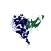
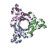
 Homo sapiens (human)
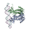
 Herpes simplex virus
 X-RAY DIFFRACTION / Resolution: 2.92 Å
  Authors
 Canada, 1items
  Citation
 Structure visualization
 Downloads & links
 Other downloads


 PDBj
PDBj


 Assembly


 Mass: 35.453 Da / Num. of mol.: 4 / Source method: obtained synthetically / Formula: Cl
 Mass: 18.015 Da / Num. of mol.: 39 / Source method: isolated from a natural source / Formula: H2O
 Sample preparation
 Processing