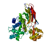
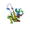
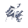
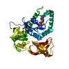
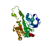
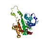
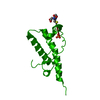
Entry Database : PDB / ID : 4wnrTitle Structure of methanosarcina barkeri Roco2 RocCORdC bound to GDP Leucine-rich-repeat protein Keywords / / / / / Function / homology Function Domain/homology Component
/ / / / / / / / / / / / / / / / / / / / / / / / / / / / / Biological species Methanosarcina barkeri (archaea)Method / / / Resolution : 2.9 Å Authors Terheyden, S. Funding support Organization Grant number Country Michael J. Fox Foundation
Journal : Biochem.J. / Year : 2015Title : Revisiting the Roco G-protein cycle.Authors : Terheyden, S. / Ho, F.Y. / Gilsbach, B.K. / Wittinghofer, A. / Kortholt, A. History Deposition Oct 14, 2014 Deposition site / Processing site Revision 1.0 Nov 5, 2014 Provider / Type Revision 1.1 Feb 11, 2015 Group Revision 1.2 Jan 10, 2024 Group Data collection / Database references ... Data collection / Database references / Derived calculations / Refinement description Category chem_comp_atom / chem_comp_bond ... chem_comp_atom / chem_comp_bond / database_2 / pdbx_initial_refinement_model / pdbx_struct_conn_angle / struct_conn Item _database_2.pdbx_DOI / _database_2.pdbx_database_accession ... _database_2.pdbx_DOI / _database_2.pdbx_database_accession / _pdbx_struct_conn_angle.ptnr1_auth_seq_id / _pdbx_struct_conn_angle.ptnr3_auth_seq_id / _pdbx_struct_conn_angle.value / _struct_conn.pdbx_dist_value / _struct_conn.ptnr2_auth_seq_id
Show all Show less
 Movie
Movie Controller


 Open data
 Basic information
 Components
 Keywords
 Function and homology information
 Methanosarcina barkeri (archaea)
 X-RAY DIFFRACTION /
  Authors
 United States, 1items
  Citation
 Structure visualization
 Downloads & links
 Other downloads

 PDBj
PDBj


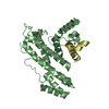
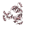
 Assembly
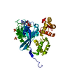


 Type: RNA linking / Mass: 443.201 Da / Num. of mol.: 1 / Source method: obtained synthetically / Formula: C10H15N5O11P2 / Comment: GDP, energy-carrying molecule*YM
 Mass: 24.305 Da / Num. of mol.: 1 / Source method: obtained synthetically / Formula: Mg
 Mass: 92.094 Da / Num. of mol.: 1 / Source method: obtained synthetically / Formula: C3H8O3
 Mass: 96.063 Da / Num. of mol.: 2 / Source method: obtained synthetically / Formula: SO4
 Sample preparation
 / Beamline: X10SA / Wavelength: 1.06997 Å
 Processing