 Movie
Movie Controller


+ Open data

- Basic information
| Entry | Database: PDB / ID: 4wjo | ||||||
|---|---|---|---|---|---|---|---|
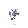
| Title | Crystal Structure of SUMO1 in complex with PML | ||||||
 Components Components |
| ||||||
 Keywords Keywords | protein binding/signaling protein / SUMO1 / PML / SUMO Interaction Motif / PhosphoSIM / protein binding-signaling protein complex | ||||||
| Function / homology |  Function and homology information Function and homology informationregulation of calcium ion transport into cytosol / ubiquitin-like protein ligase activity / negative regulation of translation in response to oxidative stress / positive regulation of protein localization to chromosome, telomeric region / PML body organization / SUMOylation of nuclear envelope proteins / SUMO is proteolytically processed / Negative regulation of activity of TFAP2 (AP-2) family transcription factors / SUMO is conjugated to E1 (UBA2:SAE1) / SUMO is transferred from E1 to E2 (UBE2I, UBC9) ...regulation of calcium ion transport into cytosol / ubiquitin-like protein ligase activity / negative regulation of translation in response to oxidative stress / positive regulation of protein localization to chromosome, telomeric region / PML body organization / SUMOylation of nuclear envelope proteins / SUMO is proteolytically processed / Negative regulation of activity of TFAP2 (AP-2) family transcription factors / SUMO is conjugated to E1 (UBA2:SAE1) / SUMO is transferred from E1 to E2 (UBE2I, UBC9) / negative regulation of transcription initiation by RNA polymerase II / fibroblast migration / negative regulation of action potential / nuclear stress granule / small protein activating enzyme binding / positive regulation of apoptotic process involved in mammary gland involution / suppression of viral release by host / SUMO binding / SUMOylation of immune response proteins / negative regulation of mitotic cell cycle / SUMOylation of SUMOylation proteins / regulation of calcium ion transmembrane transport / maintenance of protein location in nucleus / endoplasmic reticulum calcium ion homeostasis / SUMOylation of DNA methylation proteins / Dengue virus activates/modulates innate and adaptive immune responses / Maturation of nucleoprotein / XY body / oncogene-induced cell senescence / regulation of double-strand break repair / SUMOylation of RNA binding proteins / Transferases; Acyltransferases; Aminoacyltransferases / regulation of cardiac muscle cell contraction / SUMO transferase activity / Postmitotic nuclear pore complex (NPC) reformation / Regulation of RUNX1 Expression and Activity / positive regulation of extrinsic apoptotic signaling pathway / cobalt ion binding / Maturation of nucleoprotein / negative regulation of protein import into nucleus / negative regulation of interleukin-1 beta production / roof of mouth development / Maturation of DENV proteins / SUMOylation of ubiquitinylation proteins / entrainment of circadian clock by photoperiod / ubiquitin-specific protease binding / cellular response to cadmium ion / SMAD binding / negative regulation of telomere maintenance via telomerase / SUMOylation of transcription factors / SUMOylation of DNA replication proteins / positive regulation of telomere maintenance / ubiquitin-like protein ligase binding / intrinsic apoptotic signaling pathway in response to DNA damage by p53 class mediator / protein sumoylation / nuclear pore / potassium channel regulator activity / Regulation of IFNG signaling / transporter activator activity / protein targeting / postsynaptic cytosol / regulation of cell adhesion / positive regulation of signal transduction by p53 class mediator / negative regulation of ubiquitin-dependent protein catabolic process / SUMOylation of DNA damage response and repair proteins / Regulation of TP53 Activity through Acetylation / presynaptic cytosol / positive regulation of defense response to virus by host / Transcriptional and post-translational regulation of MITF-M expression and activity / response to cytokine / SUMOylation of transcription cofactors / negative regulation of angiogenesis / SUMOylation of chromatin organization proteins / Regulation of PTEN localization / DNA damage response, signal transduction by p53 class mediator / SUMOylation of intracellular receptors / circadian regulation of gene expression / establishment of protein localization / regulation of protein stability / positive regulation of protein-containing complex assembly / negative regulation of cell growth / regulation of circadian rhythm / PML body / PKR-mediated signaling / intrinsic apoptotic signaling pathway in response to DNA damage / positive regulation of fibroblast proliferation / cellular senescence / Interferon gamma signaling / Transcriptional regulation of granulopoiesis / nuclear matrix / Formation of Incision Complex in GG-NER / HCMV Early Events / protein tag activity / positive regulation of proteasomal ubiquitin-dependent protein catabolic process / cellular response to heat / Recruitment and ATM-mediated phosphorylation of repair and signaling proteins at DNA double strand breaks / protein-containing complex assembly / nuclear membrane / early endosome membrane / molecular adaptor activity Similarity search - Function | ||||||
| Biological species |  Homo sapiens (human) Homo sapiens (human) | ||||||
| Method |  X-RAY DIFFRACTION / SYNCHROTRON / MOLECULAR REPLACEMENT / Resolution: 1.46 Å X-RAY DIFFRACTION / SYNCHROTRON / MOLECULAR REPLACEMENT / Resolution: 1.46 Å | ||||||
 Authors Authors | Cappadocia, L. / Mascle, X.H. / Bourdeau, V. / Tremblay-Belzile, S. / Chaker-Margot, M. / Lussier-Price, M. / Wada, J. / Sakaguchi, K. / Aubry, M. / Ferbeyre, G. / Omichinski, J.G. | ||||||
 Citation Citation | Journal: Structure / Year: 2015 Title: Structural and Functional Characterization of the Phosphorylation-Dependent Interaction between PML and SUMO1. Authors: Cappadocia, L. / Mascle, X.H. / Bourdeau, V. / Tremblay-Belzile, S. / Chaker-Margot, M. / Lussier-Price, M. / Wada, J. / Sakaguchi, K. / Aubry, M. / Ferbeyre, G. / Omichinski, J.G. | ||||||
| History |
|
- Structure visualization
| Structure viewer | Molecule: MolmilJmol/JSmol |
|---|
- Downloads & links
-Download
| PDBx/mmCIF format | 4wjo.cif.gz | 71.8 KB | Display | PDBx/mmCIF format |
|---|---|---|---|---|
| PDB format | pdb4wjo.ent.gz | 52.6 KB | Display | PDB format |
| PDBx/mmJSON format | 4wjo.json.gz | Tree view | PDBx/mmJSON format | |
| Others |  Other downloads Other downloads |
-Validation report
| Arichive directory | https://data.pdbj.org/pub/pdb/validation_reports/wj/4wjoftp://data.pdbj.org/pub/pdb/validation_reports/wj/4wjo | HTTPS FTP |
|---|
-Related structure data
| Related structure data |  4wjnC  4wjpC  4wjqC 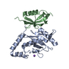 2uyzS C: citing same article ( S: Starting model for refinement |
|---|---|
| Similar structure data |


-Links
 PDBj
PDBj


- Assembly
| Deposited unit | 
| ||||||||
|---|---|---|---|---|---|---|---|---|---|
| 1 |
| ||||||||
| Unit cell |
|
-Components
| #1: Protein | Mass: 9526.771 Da / Num. of mol.: 1 / Fragment: SUMO1, unp residues 17-97 / Mutation: C52A Source method: isolated from a genetically manipulated source Source: (gene. exp.) Homo sapiens (human) / Gene: SUMO1, SMT3C, SMT3H3, UBL1, OK/SW-cl.43 / Plasmid: pGEX2T / Production host:  |
|---|---|
| #2: Protein/peptide | Mass: 2976.985 Da / Num. of mol.: 1 / Fragment: PML, unp residues 547-573 Source method: isolated from a genetically manipulated source Source: (gene. exp.) Homo sapiens (human) / Gene: PML, MYL, PP8675, RNF71, TRIM19 / Plasmid: pGEX2T / Production host: |
| #3: Water | ChemComp-HOH /  Mass: 18.015 Da / Num. of mol.: 148 / Source method: isolated from a natural source / Formula: H2O Mass: 18.015 Da / Num. of mol.: 148 / Source method: isolated from a natural source / Formula: H2O |
-Experimental details
-Experiment
| Experiment | Method: X-RAY DIFFRACTION / Number of used crystals: 1 |
|---|
- Sample preparation
| Crystal | Density Matthews: 2.31 Å3/Da / Density % sol: 46.8 % |
|---|---|
| Crystal grow | Temperature: 298 K / Method: vapor diffusion, hanging drop / pH: 6.5 Details: 100mM sodium cacodylate pH6.5, 16% PEG3350, 10mM calcium chloride |
-Data collection
| Diffraction | Mean temperature: 100 K |
|---|---|
| Diffraction source | Source: SYNCHROTRON / Site: NSLS  / Beamline: X29A / Wavelength: 0.98 Å / Beamline: X29A / Wavelength: 0.98 Å |
| Detector | Type: ADSC QUANTUM 315 / Detector: CCD / Date: Oct 11, 2012 |
| Radiation | Monochromator: DOUBLE CRYSTAL MONOCHROMATOR / Protocol: SINGLE WAVELENGTH / Monochromatic (M) / Laue (L): M / Scattering type: x-ray |
| Radiation wavelength | Wavelength: 0.98 Å / Relative weight: 1 |
| Reflection | Resolution: 1.46→50 Å / Num. obs: 20770 / % possible obs: 99.2 % / Redundancy: 6.8 % / Biso Wilson estimate: 14.96 Å2 / Rmerge(I) obs: 0.092 / Net I/σ(I): 11.6 |
| Reflection shell | Resolution: 1.46→1.54 Å / Redundancy: 5.6 % / Rmerge(I) obs: 0.597 / Mean I/σ(I) obs: 2.7 / % possible all: 95.4 |
- Processing
| Software |
| ||||||||||||||||||||||||||||||||||||||||||||||||||||||||||||||||||||||||||||||||||||||||||||||||||||
|---|---|---|---|---|---|---|---|---|---|---|---|---|---|---|---|---|---|---|---|---|---|---|---|---|---|---|---|---|---|---|---|---|---|---|---|---|---|---|---|---|---|---|---|---|---|---|---|---|---|---|---|---|---|---|---|---|---|---|---|---|---|---|---|---|---|---|---|---|---|---|---|---|---|---|---|---|---|---|---|---|---|---|---|---|---|---|---|---|---|---|---|---|---|---|---|---|---|---|---|---|---|
| Refinement | Method to determine structure: MOLECULAR REPLACEMENT Starting model: PDB ENTRY 2UYZ Resolution: 1.46→26.47 Å / FOM work R set: 0.8933 / SU ML: 0.11 / Cross valid method: FREE R-VALUE / σ(F): 1.34 / Phase error: 16.94 / Stereochemistry target values: ML
| ||||||||||||||||||||||||||||||||||||||||||||||||||||||||||||||||||||||||||||||||||||||||||||||||||||
| Solvent computation | Shrinkage radii: 0.9 Å / VDW probe radii: 1.11 Å / Solvent model: FLAT BULK SOLVENT MODEL | ||||||||||||||||||||||||||||||||||||||||||||||||||||||||||||||||||||||||||||||||||||||||||||||||||||
| Displacement parameters | Biso max: 101.03 Å2 / Biso mean: 21.56 Å2 / Biso min: 8.82 Å2 | ||||||||||||||||||||||||||||||||||||||||||||||||||||||||||||||||||||||||||||||||||||||||||||||||||||
| Refinement step | Cycle: final / Resolution: 1.46→26.47 Å
| ||||||||||||||||||||||||||||||||||||||||||||||||||||||||||||||||||||||||||||||||||||||||||||||||||||
| Refine LS restraints |
| ||||||||||||||||||||||||||||||||||||||||||||||||||||||||||||||||||||||||||||||||||||||||||||||||||||
| LS refinement shell | Refine-ID: X-RAY DIFFRACTION / Total num. of bins used: 7
| ||||||||||||||||||||||||||||||||||||||||||||||||||||||||||||||||||||||||||||||||||||||||||||||||||||
| Refinement TLS params. | Method: refined / Refine-ID: X-RAY DIFFRACTION
| ||||||||||||||||||||||||||||||||||||||||||||||||||||||||||||||||||||||||||||||||||||||||||||||||||||
| Refinement TLS group |
|
