 Movie
Movie Controller

[English] 日本語
 Yorodumi
- PDB-4wid: Crystal structure of the immediate-early 1 protein (IE1) at 2.31 ... -
+ Open data

- Basic information
| Entry | Database: PDB / ID: 4wid | |||||||||
|---|---|---|---|---|---|---|---|---|---|---|
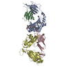
| Title | Crystal structure of the immediate-early 1 protein (IE1) at 2.31 angstrom (tetragonal form after crystal dehydration) | |||||||||
 Components Components | (RhUL123) x 2 | |||||||||
 Keywords Keywords | VIRAL PROTEIN / Antagonist / cytomegalovirus | |||||||||
| Function / homology | Cytomegalovirus IE1/IE2 / Cytomegalovirus IE1 protein / DNA-templated viral transcription / symbiont-mediated perturbation of host cell cycle G1/S transition checkpoint / host cell nucleus / RhUL123 Function and homology information Function and homology information | |||||||||
| Biological species |  Macacine herpesvirus 3 (Rhesus cytomegalovirus) Macacine herpesvirus 3 (Rhesus cytomegalovirus) | |||||||||
| Method |  X-RAY DIFFRACTION / SYNCHROTRON / MOLECULAR REPLACEMENT / Resolution: 2.31 Å X-RAY DIFFRACTION / SYNCHROTRON / MOLECULAR REPLACEMENT / Resolution: 2.31 Å | |||||||||
 Authors Authors | Klingl, S. / Scherer, M. / Sevvana, M. / Muller, Y.A. / Stamminger, T. | |||||||||
| Funding support |  Germany, 1items Germany, 1items
| |||||||||
 Citation Citation | Journal: Plos Pathog. / Year: 2014 Title: Crystal Structure of Cytomegalovirus IE1 Protein Reveals Targeting of TRIM Family Member PML via Coiled-Coil Interactions. Authors: Scherer, M. / Klingl, S. / Sevvana, M. / Otto, V. / Schilling, E.M. / Stump, J.D. / Muller, R. / Reuter, N. / Sticht, H. / Muller, Y.A. / Stamminger, T. | |||||||||
| History |
|
- Structure visualization
| Structure viewer | Molecule: MolmilJmol/JSmol |
|---|
- Downloads & links
-Download
| PDBx/mmCIF format | 4wid.cif.gz | 300.3 KB | Display | PDBx/mmCIF format |
|---|---|---|---|---|
| PDB format | pdb4wid.ent.gz | 244.3 KB | Display | PDB format |
| PDBx/mmJSON format | 4wid.json.gz | Tree view | PDBx/mmJSON format | |
| Others |  Other downloads Other downloads |
-Validation report
| Arichive directory | https://data.pdbj.org/pub/pdb/validation_reports/wi/4widftp://data.pdbj.org/pub/pdb/validation_reports/wi/4wid | HTTPS FTP |
|---|
-Related structure data
| Related structure data |  4w1cS S: Starting model for refinement |
|---|---|
| Similar structure data |


-Links
 PDBj
PDBj- Assembly
| Deposited unit | 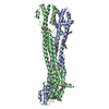
| ||||||||
|---|---|---|---|---|---|---|---|---|---|
| 1 |
| ||||||||
| Unit cell |
|
-Components
| #1: Protein | Mass: 42265.668 Da / Num. of mol.: 1 / Fragment: UNP residues 36-395 Source method: isolated from a genetically manipulated source Source: (gene. exp.) Macacine herpesvirus 3 (Rhesus cytomegalovirus)Plasmid: pGEX-6P-1 / Production host:  | ||
|---|---|---|---|
| #2: Protein | Mass: 42239.633 Da / Num. of mol.: 1 / Fragment: UNP residues 36-395 Source method: isolated from a genetically manipulated source Source: (gene. exp.) Macacine herpesvirus 3 (Rhesus cytomegalovirus)Plasmid: pGEX-6P-1 / Production host: | ||
| #3: Chemical | ChemComp-TRS /   Mass: 122.143 Da / Num. of mol.: 1 / Source method: obtained synthetically / Formula: C4H12NO3 / Comment: pH buffer*YM Mass: 122.143 Da / Num. of mol.: 1 / Source method: obtained synthetically / Formula: C4H12NO3 / Comment: pH buffer*YM | ||
| #4: Chemical |   Mass: 62.068 Da / Num. of mol.: 2 / Source method: obtained synthetically / Formula: C2H6O2 Mass: 62.068 Da / Num. of mol.: 2 / Source method: obtained synthetically / Formula: C2H6O2#5: Water | ChemComp-HOH / |  Mass: 18.015 Da / Num. of mol.: 123 / Source method: isolated from a natural source / Formula: H2O Mass: 18.015 Da / Num. of mol.: 123 / Source method: isolated from a natural source / Formula: H2O |
-Experimental details
-Experiment
| Experiment | Method: X-RAY DIFFRACTION |
|---|
- Sample preparation
| Crystal | Density Matthews: 2.62 Å3/Da / Density % sol: 53.16 % |
|---|---|
| Crystal grow | Temperature: 293 K / Method: vapor diffusion, hanging drop Details: Reservoir 700 microL: 15 % (w/v) PEG 3350, 400 mM magnesium formate, Drop ratio: 1 to 2 microL protein solution (20 mg/mL) plus 1 microL of reservoir solution supplemented with crystal microseeds PH range: approx. 7.0 |
-Data collection
| Diffraction | Mean temperature: 100 K |
|---|---|
| Diffraction source | Source: SYNCHROTRON / Site: BESSY / Beamline: 14.1 / Wavelength: 0.9184 Å |
| Detector | Type: MARMOSAIC 225 mm CCD / Detector: CCD / Date: Dec 17, 2011 |
| Radiation | Protocol: SINGLE WAVELENGTH / Monochromatic (M) / Laue (L): M / Scattering type: x-ray |
| Radiation wavelength | Wavelength: 0.9184 Å / Relative weight: 1 |
| Reflection | Resolution: 2.31→48.18 Å / Num. obs: 37710 / % possible obs: 99.27 % / Redundancy: 5.9 % / Rmerge(I) obs: 0.059 / Net I/σ(I): 16.52 |
| Reflection shell | Resolution: 2.306→2.389 Å / Redundancy: 3.3 % / Rmerge(I) obs: 0.59 / Mean I/σ(I) obs: 1.56 / % possible all: 93.66 |
- Processing
| Software |
| |||||||||||||||||||||||||||||||||||||||||||||||||||||||||||||||||||||||||||||||||||||||||||||||||||||||||
|---|---|---|---|---|---|---|---|---|---|---|---|---|---|---|---|---|---|---|---|---|---|---|---|---|---|---|---|---|---|---|---|---|---|---|---|---|---|---|---|---|---|---|---|---|---|---|---|---|---|---|---|---|---|---|---|---|---|---|---|---|---|---|---|---|---|---|---|---|---|---|---|---|---|---|---|---|---|---|---|---|---|---|---|---|---|---|---|---|---|---|---|---|---|---|---|---|---|---|---|---|---|---|---|---|---|---|
| Refinement | Method to determine structure: MOLECULAR REPLACEMENT Starting model: Chain A of PDB 4W1C Resolution: 2.31→48.18 Å / SU ML: 0.35 / Cross valid method: FREE R-VALUE / σ(F): 2.01 / Phase error: 31.19 / Stereochemistry target values: ML
| |||||||||||||||||||||||||||||||||||||||||||||||||||||||||||||||||||||||||||||||||||||||||||||||||||||||||
| Solvent computation | Shrinkage radii: 0.9 Å / VDW probe radii: 1.11 Å / Solvent model: FLAT BULK SOLVENT MODEL | |||||||||||||||||||||||||||||||||||||||||||||||||||||||||||||||||||||||||||||||||||||||||||||||||||||||||
| Refinement step | Cycle: LAST / Resolution: 2.31→48.18 Å
| |||||||||||||||||||||||||||||||||||||||||||||||||||||||||||||||||||||||||||||||||||||||||||||||||||||||||
| Refine LS restraints |
| |||||||||||||||||||||||||||||||||||||||||||||||||||||||||||||||||||||||||||||||||||||||||||||||||||||||||
| LS refinement shell |
| |||||||||||||||||||||||||||||||||||||||||||||||||||||||||||||||||||||||||||||||||||||||||||||||||||||||||
| Refinement TLS params. | Method: refined / Refine-ID: X-RAY DIFFRACTION
| |||||||||||||||||||||||||||||||||||||||||||||||||||||||||||||||||||||||||||||||||||||||||||||||||||||||||
| Refinement TLS group |
|
