| Entry | Database: PDB / ID: 4whf
|
|---|
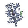
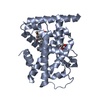
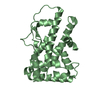
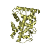
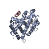
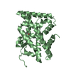
| Title | Crystal Structure of TR3 LBD in complex with 1-(3,4,5-trihydroxyphenyl)decan-1-one |
|---|
 Components Components | Nuclear receptor subfamily 4 group A member 1 |
|---|
 Keywords Keywords | TRANSCRIPTION / LBD |
|---|
| Function / homology |  Function and homology information Function and homology information
cellular response to corticotropin-releasing hormone stimulus / regulation of type B pancreatic cell proliferation / non-canonical inflammasome complex assembly / detection of lipopolysaccharide / AKT phosphorylates targets in the nucleus / endothelial cell chemotaxis / nuclear glucocorticoid receptor binding / macrophage activation / cell migration involved in sprouting angiogenesis / Constitutive Signaling by AKT1 E17K in Cancer ...cellular response to corticotropin-releasing hormone stimulus / regulation of type B pancreatic cell proliferation / non-canonical inflammasome complex assembly / detection of lipopolysaccharide / AKT phosphorylates targets in the nucleus / endothelial cell chemotaxis / nuclear glucocorticoid receptor binding / macrophage activation / cell migration involved in sprouting angiogenesis / Constitutive Signaling by AKT1 E17K in Cancer / negative regulation of cell cycle / cellular response to vascular endothelial growth factor stimulus / skeletal muscle cell differentiation / fat cell differentiation / cellular response to fibroblast growth factor stimulus / positive regulation of endothelial cell proliferation / lipopolysaccharide binding / Nuclear Receptor transcription pathway / nuclear receptor activity / fibrillar center / protein import into nucleus / sequence-specific double-stranded DNA binding / DNA-binding transcription activator activity, RNA polymerase II-specific / transcription regulator complex / nuclear membrane / transcription by RNA polymerase II / DNA-binding transcription factor activity, RNA polymerase II-specific / nuclear speck / RNA polymerase II cis-regulatory region sequence-specific DNA binding / positive regulation of apoptotic process / protein heterodimerization activity / inflammatory response / apoptotic process / regulation of transcription by RNA polymerase II / chromatin / signal transduction / positive regulation of transcription by RNA polymerase II / mitochondrion / DNA binding / zinc ion binding / nucleoplasm / identical protein binding / nucleus / cytosolSimilarity search - Function Orphan nuclear receptor, HMR type / Orphan nuclear receptor / Retinoid X Receptor / Retinoid X Receptor / Nuclear hormone receptor / Nuclear hormones receptors DNA-binding region signature. / Zinc finger, nuclear hormone receptor-type / Double treble clef zinc finger, C4 type / Nuclear hormone receptors DNA-binding domain profile. / c4 zinc finger in nuclear hormone receptors ...Orphan nuclear receptor, HMR type / Orphan nuclear receptor / Retinoid X Receptor / Retinoid X Receptor / Nuclear hormone receptor / Nuclear hormones receptors DNA-binding region signature. / Zinc finger, nuclear hormone receptor-type / Double treble clef zinc finger, C4 type / Nuclear hormone receptors DNA-binding domain profile. / c4 zinc finger in nuclear hormone receptors / Nuclear hormone receptor, ligand-binding domain / Nuclear hormone receptor-like domain superfamily / Ligand-binding domain of nuclear hormone receptor / Nuclear receptor (NR) ligand-binding (LBD) domain profile. / Ligand binding domain of hormone receptors / Zinc finger, NHR/GATA-type / Orthogonal Bundle / Mainly AlphaSimilarity search - Domain/homology |
|---|
| Biological species |  Homo sapiens (human) Homo sapiens (human) |
|---|
| Method |  X-RAY DIFFRACTION / SYNCHROTRON / MOLECULAR REPLACEMENT / molecular replacement / Resolution: 2.27 Å X-RAY DIFFRACTION / SYNCHROTRON / MOLECULAR REPLACEMENT / molecular replacement / Resolution: 2.27 Å |
|---|
 Authors Authors | Li, F.W. / Cai, Q.X. / Li, A.Z. / Tian, X.Y. / Wang, W.J. / Wang, Y. / Hou, P.P. / Wu, Q. / Lin, T.W. |
|---|
 Citation Citation | Journal: Chem.Biol. / Year: 2015
Title: Induction of Autophagic Death in Cancer Cells by Agonizing TR3 and Attenuating Akt2 Activity
Authors: Wang, W.J. / Wang, Y. / Hou, P.P. / Li, F.W. / Zhou, B. / Chen, H.Z. / Bian, X.L. / Cai, Q.X. / Xing, Y.Z. / He, J.P. / Zhang, H. / Huang, P.Q. / Lin, T. / Wu, Q. |
|---|
| History | | Deposition | Sep 22, 2014 | Deposition site: RCSB / Processing site: PDBJ |
|---|
| Revision 1.0 | Sep 9, 2015 | Provider: repository / Type: Initial release |
|---|
| Revision 1.1 | Oct 14, 2015 | Group: Derived calculations |
|---|
| Revision 1.2 | Mar 20, 2024 | Group: Data collection / Database references ...Data collection / Database references / Derived calculations / Refinement description
Category: chem_comp_atom / chem_comp_bond ...chem_comp_atom / chem_comp_bond / database_2 / pdbx_struct_oper_list / struct_ncs_dom_lim
Item: _database_2.pdbx_DOI / _database_2.pdbx_database_accession ..._database_2.pdbx_DOI / _database_2.pdbx_database_accession / _pdbx_struct_oper_list.symmetry_operation / _struct_ncs_dom_lim.beg_auth_comp_id / _struct_ncs_dom_lim.beg_label_asym_id / _struct_ncs_dom_lim.beg_label_comp_id / _struct_ncs_dom_lim.beg_label_seq_id / _struct_ncs_dom_lim.end_auth_comp_id / _struct_ncs_dom_lim.end_label_asym_id / _struct_ncs_dom_lim.end_label_comp_id / _struct_ncs_dom_lim.end_label_seq_id |
|---|
|
|---|
 Movie
Movie Controller

 Yorodumi
 Open data
 Basic information
 Structure visualization
 Downloads & links
 Other downloads


 PDBj
PDBj


 Assembly
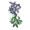


 Mass: 92.094 Da / Num. of mol.: 5 / Source method: obtained synthetically / Formula: C3H8O3
 Mass: 280.359 Da / Num. of mol.: 1 / Source method: obtained synthetically / Formula: C16H24O4
 Mass: 18.015 Da / Num. of mol.: 190 / Source method: isolated from a natural source / Formula: H2O
 Sample preparation
 / Beamline: BL17U / Wavelength: 0.98 Å
 Processing