- PDB-4wed: Crystal structure of ABC transporter substrate-binding protein fr... -
+
Open data
ID or keywords:
Loading...
-
Basic information
Entry
Database: PDB / ID: 4wed
Title
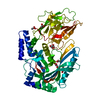
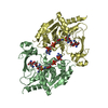
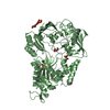
Crystal structure of ABC transporter substrate-binding protein from Sinorhizobium meliloti
Components
ABC transporter, periplasmic solute-binding protein
Keywords
TRANSPORT PROTEIN / ABC transporter substrate-binding protein / Sinorhizobium meliloti / Structural Genomics / NYSGRC / PSI-Biology / New York Structural Genomics Research Consortium
Function / homology
Function and homology information
nickel cation transport / peptide transport / peptide transmembrane transporter activity / nickel cation binding / ATP-binding cassette (ABC) transporter complex / outer membrane-bounded periplasmic space / heme binding Similarity search - Function
Nickel ABC transporter, substrate-binding protein NikA / Solute-binding protein family 5, conserved site / Bacterial extracellular solute-binding proteins, family 5 signature. / Dipeptide-binding Protein; domain 3 / Dipeptide-binding Protein; Domain 3 / Peptide/nickel binding protein, MppA-type / Solute-binding protein family 5 domain / Solute-binding protein family 5 / Bacterial extracellular solute-binding proteins, family 5 Middle / Twin arginine translocation (Tat) signal profile. ...Nickel ABC transporter, substrate-binding protein NikA / Solute-binding protein family 5, conserved site / Bacterial extracellular solute-binding proteins, family 5 signature. / Dipeptide-binding Protein; domain 3 / Dipeptide-binding Protein; Domain 3 / Peptide/nickel binding protein, MppA-type / Solute-binding protein family 5 domain / Solute-binding protein family 5 / Bacterial extracellular solute-binding proteins, family 5 Middle / Twin arginine translocation (Tat) signal profile. / Twin-arginine translocation pathway, signal sequence / Periplasmic binding protein-like II / D-Maltodextrin-Binding Protein; domain 2 / Roll / 3-Layer(aba) Sandwich / Alpha Beta Similarity search - Domain/homology
Mass: 18.015 Da / Num. of mol.: 101 / Source method: isolated from a natural source / Formula: H2O
Has protein modification
Y
Sequence details
There was an additional tag present in the crystallization setup, but it was likely cleaved by the ...There was an additional tag present in the crystallization setup, but it was likely cleaved by the chymotrypsin present. The sequence was MHHHHHHSSGVDLGTENLYFQS
-
Experimental details
-
Experiment
Experiment
Method: X-RAY DIFFRACTION / Number of used crystals: 1
-
Sample preparation
Crystal
Density Matthews: 2.18 Å3/Da / Density % sol: 43.54 %
Crystal grow
Temperature: 289 K / Method: vapor diffusion, sitting drop / pH: 7.5 Details: 0.2 ul of 18 mg/ml protein in 20 mM HEPES pH 7.5, 150 mM NaCl, 10% Glycerol, 0.1% Sodium Azide and 0.5 mM TCEP were mixed with 0.2 ul of the MCSG-II condition # 18 (0.2 M Sodium Formate, 20% ...Details: 0.2 ul of 18 mg/ml protein in 20 mM HEPES pH 7.5, 150 mM NaCl, 10% Glycerol, 0.1% Sodium Azide and 0.5 mM TCEP were mixed with 0.2 ul of the MCSG-II condition # 18 (0.2 M Sodium Formate, 20% (w/v) PEG 3350 ) and equilibrated against 2.0 M NaCl solution in 96 Well 3 drop Crystallization Plate (Swissci). Before crystallization protein was incubated with 1/50 v/v of 2 mg/ml chymotrypsin solution at 289 K for 3 hours.
Method to determine structure: SAD / Resolution: 2.35→49.65 Å / Cor.coef. Fo:Fc: 0.97 / Cor.coef. Fo:Fc free: 0.952 / WRfactor Rfree: 0.249 / WRfactor Rwork: 0.1741 / FOM work R set: 0.8026 / SU B: 18.319 / SU ML: 0.21 / SU R Cruickshank DPI: 0.383 / SU Rfree: 0.2455 / Cross valid method: THROUGHOUT / σ(F): 0 / ESU R: 0.383 / ESU R Free: 0.245 / Stereochemistry target values: MAXIMUM LIKELIHOOD Details: U VALUES : WITH TLS ADDED HYDROGENS HAVE BEEN ADDED IN THE RIDING POSITIONS
Rfactor
Num. reflection
% reflection
Selection details
Rfree
0.2294
899
4.6 %
RANDOM
Rwork
0.1641
18838
-
-
obs
0.1667
-
97.39 %
-
Solvent computation
Ion probe radii: 0.8 Å / Shrinkage radii: 0.8 Å / VDW probe radii: 1.2 Å / Solvent model: BABINET MODEL WITH MASK
In the structure databanks used in Yorodumi, some data are registered as the other names, "COVID-19 virus" and "2019-nCoV". Here are the details of the virus and the list of structure data.
Jan 31, 2019. EMDB accession codes are about to change! (news from PDBe EMDB page)
EMDB accession codes are about to change! (news from PDBe EMDB page)
The allocation of 4 digits for EMDB accession codes will soon come to an end. Whilst these codes will remain in use, new EMDB accession codes will include an additional digit and will expand incrementally as the available range of codes is exhausted. The current 4-digit format prefixed with “EMD-” (i.e. EMD-XXXX) will advance to a 5-digit format (i.e. EMD-XXXXX), and so on. It is currently estimated that the 4-digit codes will be depleted around Spring 2019, at which point the 5-digit format will come into force.
The EM Navigator/Yorodumi systems omit the EMD- prefix.
Related info.:Q: What is EMD? / ID/Accession-code notation in Yorodumi/EM Navigator
Yorodumi is a browser for structure data from EMDB, PDB, SASBDB, etc.
This page is also the successor to EM Navigator detail page, and also detail information page/front-end page for Omokage search.
The word "yorodu" (or yorozu) is an old Japanese word meaning "ten thousand". "mi" (miru) is to see.
Related info.:EMDB / PDB / SASBDB / Comparison of 3 databanks / Yorodumi Search / Aug 31, 2016. New EM Navigator & Yorodumi / Yorodumi Papers / Jmol/JSmol / Function and homology information / Changes in new EM Navigator and Yorodumi
 Movie
Movie Controller

 Yorodumi
 Open data
 Basic information
 Components
 Keywords
 Function and homology information
 Rhizobium meliloti (bacteria)
 X-RAY DIFFRACTION /
  Authors
 United States, 1items
  Citation
 Structure visualization
 Downloads & links
 Other downloads
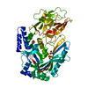


 PDBj
PDBj

 Assembly
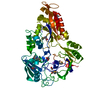

 Mass: 22.990 Da / Num. of mol.: 1 / Source method: obtained synthetically / Formula: Na
 Mass: 46.025 Da / Num. of mol.: 1 / Source method: obtained synthetically / Formula: CH2O2
 Mass: 92.094 Da / Num. of mol.: 1 / Source method: obtained synthetically / Formula: C3H8O3
 Mass: 18.015 Da / Num. of mol.: 101 / Source method: isolated from a natural source / Formula: H2O
 Sample preparation
 Processing