 Movie
Movie Controller


+ Open data

- Basic information
| Entry | Database: PDB / ID: 4wbh | ||||||
|---|---|---|---|---|---|---|---|
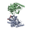
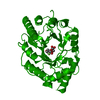
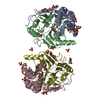
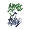
| Title | STRUCTURE OF THE WNT DEACYLASE NOTUM - CRYSTAL FORM I APO - 2.2A | ||||||
 Components Components | Protein notum homolog | ||||||
 Keywords Keywords | HYDROLASE / Wnt / extracellular Esterase / alpha/beta hydrolase | ||||||
| Function / homology |  Function and homology information Function and homology information[Wnt protein] O-palmitoleoyl-L-serine hydrolase / protein-containing complex destabilizing activity / protein depalmitoleylation / palmitoleyl hydrolase activity / Release of Hh-Np from the secreting cell / C-type glycerophospholipase activity / regulation of bone mineralization / negative regulation of Wnt signaling pathway / Post-translational protein phosphorylation / negative regulation of canonical Wnt signaling pathway ...[Wnt protein] O-palmitoleoyl-L-serine hydrolase / protein-containing complex destabilizing activity / protein depalmitoleylation / palmitoleyl hydrolase activity / Release of Hh-Np from the secreting cell / C-type glycerophospholipase activity / regulation of bone mineralization / negative regulation of Wnt signaling pathway / Post-translational protein phosphorylation / negative regulation of canonical Wnt signaling pathway / bone development / Regulation of Insulin-like Growth Factor (IGF) transport and uptake by Insulin-like Growth Factor Binding Proteins (IGFBPs) / Wnt signaling pathway / endoplasmic reticulum lumen / extracellular region Similarity search - Function | ||||||
| Biological species |  Homo sapiens (human) Homo sapiens (human) | ||||||
| Method |  X-RAY DIFFRACTION / SYNCHROTRON / SAD / Resolution: 2.2 Å X-RAY DIFFRACTION / SYNCHROTRON / SAD / Resolution: 2.2 Å | ||||||
 Authors Authors | Zebisch, M. / Jones, E.Y. | ||||||
| Funding support |  United Kingdom, 1items United Kingdom, 1items
| ||||||
 Citation Citation | Journal: Nature / Year: 2015 Title: Notum deacylates Wnt proteins to suppress signalling activity Authors: Kakugawa, S. / Langton, P.F. / Zebisch, M. / Howell, S.A. / Chang, T.-H. / Liu, Y. / Feizi, T. / Bineva, G. / O'Reilly, N. / Snijders, A.P. / Jones, E.Y. / Vincent, J.-P. | ||||||
| History |
|
- Structure visualization
| Structure viewer | Molecule: MolmilJmol/JSmol |
|---|
- Downloads & links
-Download
| PDBx/mmCIF format | 4wbh.cif.gz | 292.6 KB | Display | PDBx/mmCIF format |
|---|---|---|---|---|
| PDB format | pdb4wbh.ent.gz | 236.1 KB | Display | PDB format |
| PDBx/mmJSON format | 4wbh.json.gz | Tree view | PDBx/mmJSON format | |
| Others |  Other downloads Other downloads |
-Validation report
| Arichive directory | https://data.pdbj.org/pub/pdb/validation_reports/wb/4wbhftp://data.pdbj.org/pub/pdb/validation_reports/wb/4wbh | HTTPS FTP |
|---|
-Related structure data
| Related structure data |  4uyuC  4uywC 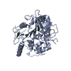 4uyzC  4uz1C  4uz5C 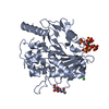 4uz6C 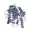 4uz7C 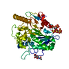 4uz9C  4uzaC  4uzjC  4uzkC  4uzlC  4uzqC C: citing same article ( |
|---|---|
| Similar structure data |


-Links
 PDBj
PDBj- Assembly
| Deposited unit | 
| ||||||||
|---|---|---|---|---|---|---|---|---|---|
| 1 |
| ||||||||
| Unit cell |
|
-Components
| #1: Protein | Mass: 53098.891 Da / Num. of mol.: 2 / Fragment: UNP residues 38-496 Source method: isolated from a genetically manipulated source Source: (gene. exp.) Homo sapiens (human) / Gene: NOTUM, OK/SW-CL.30 / Plasmid: pHLsec / Cell line (production host): HEK293T / Production host: Homo sapiens (human) / References: UniProt: Q6P988, Hydrolases#2: Chemical | ChemComp-CL /   Mass: 35.453 Da / Num. of mol.: 4 / Source method: obtained synthetically / Formula: Cl Mass: 35.453 Da / Num. of mol.: 4 / Source method: obtained synthetically / Formula: Cl#3: Sugar | ChemComp-NAG / |   Type: D-saccharide, beta linking / Mass: 221.208 Da / Num. of mol.: 1 Type: D-saccharide, beta linking / Mass: 221.208 Da / Num. of mol.: 1Source method: isolated from a genetically manipulated source Formula: C8H15NO6 #4: Chemical | ChemComp-SO4 / |   Mass: 96.063 Da / Num. of mol.: 1 / Source method: obtained synthetically / Formula: SO4 Mass: 96.063 Da / Num. of mol.: 1 / Source method: obtained synthetically / Formula: SO4#5: Water | ChemComp-HOH / |  Mass: 18.015 Da / Num. of mol.: 59 / Source method: isolated from a natural source / Formula: H2O Mass: 18.015 Da / Num. of mol.: 59 / Source method: isolated from a natural source / Formula: H2OHas protein modification | Y | |
|---|
-Experimental details
-Experiment
| Experiment | Method: X-RAY DIFFRACTION |
|---|
- Sample preparation
| Crystal | Density Matthews: 1.82 Å3/Da / Density % sol: 32.58 % |
|---|---|
| Crystal grow | Temperature: 293 K / Method: vapor diffusion / pH: 6 Details: 30% PEG3350, 100mM BisTris 6.0, 5% Glycerol, 200mM Ammonium Sulfate |
-Data collection
| Diffraction | Mean temperature: 100 K |
|---|---|
| Diffraction source | Source: SYNCHROTRON / Site: Diamond / Beamline: I04 / Wavelength: 0.9795 Å |
| Detector | Type: ADSC QUANTUM 270 / Detector: CCD / Date: Apr 25, 2012 |
| Radiation | Protocol: SINGLE WAVELENGTH / Monochromatic (M) / Laue (L): M / Scattering type: x-ray |
| Radiation wavelength | Wavelength: 0.9795 Å / Relative weight: 1 |
| Reflection | Resolution: 2.2→67 Å / Num. obs: 40214 / % possible obs: 99.7 % / Redundancy: 4 % / Rmerge(I) obs: 0.08 / Net I/σ(I): 10.1 |
| Reflection shell | Resolution: 2.2→2.32 Å / Redundancy: 4.1 % / Rmerge(I) obs: 0.608 / Mean I/σ(I) obs: 1.9 / Num. unique all: 4790 / % possible all: 99 |
- Processing
| Software |
| ||||||||||||||||||||||||||||||||||||||||||||||||||||||||||||||||||||||||||||||||||||||||||||||||||||||||||||||||||||||||||||||||||||||||||||||||||||||||||||||||||||||||||||||||||||||
|---|---|---|---|---|---|---|---|---|---|---|---|---|---|---|---|---|---|---|---|---|---|---|---|---|---|---|---|---|---|---|---|---|---|---|---|---|---|---|---|---|---|---|---|---|---|---|---|---|---|---|---|---|---|---|---|---|---|---|---|---|---|---|---|---|---|---|---|---|---|---|---|---|---|---|---|---|---|---|---|---|---|---|---|---|---|---|---|---|---|---|---|---|---|---|---|---|---|---|---|---|---|---|---|---|---|---|---|---|---|---|---|---|---|---|---|---|---|---|---|---|---|---|---|---|---|---|---|---|---|---|---|---|---|---|---|---|---|---|---|---|---|---|---|---|---|---|---|---|---|---|---|---|---|---|---|---|---|---|---|---|---|---|---|---|---|---|---|---|---|---|---|---|---|---|---|---|---|---|---|---|---|---|---|
| Refinement | Method to determine structure: SAD / Resolution: 2.2→87.72 Å / Cor.coef. Fo:Fc: 0.937 / Cor.coef. Fo:Fc free: 0.907 / SU B: 16.789 / SU ML: 0.186 / Cross valid method: THROUGHOUT / ESU R: 0.291 / ESU R Free: 0.226 / Stereochemistry target values: MAXIMUM LIKELIHOOD / Details: HYDROGENS HAVE BEEN USED IF PRESENT IN THE INPUT
| ||||||||||||||||||||||||||||||||||||||||||||||||||||||||||||||||||||||||||||||||||||||||||||||||||||||||||||||||||||||||||||||||||||||||||||||||||||||||||||||||||||||||||||||||||||||
| Solvent computation | Ion probe radii: 0.8 Å / Shrinkage radii: 0.8 Å / VDW probe radii: 1.2 Å / Solvent model: MASK | ||||||||||||||||||||||||||||||||||||||||||||||||||||||||||||||||||||||||||||||||||||||||||||||||||||||||||||||||||||||||||||||||||||||||||||||||||||||||||||||||||||||||||||||||||||||
| Displacement parameters | Biso mean: 43.707 Å2
| ||||||||||||||||||||||||||||||||||||||||||||||||||||||||||||||||||||||||||||||||||||||||||||||||||||||||||||||||||||||||||||||||||||||||||||||||||||||||||||||||||||||||||||||||||||||
| Refinement step | Cycle: LAST / Resolution: 2.2→87.72 Å
| ||||||||||||||||||||||||||||||||||||||||||||||||||||||||||||||||||||||||||||||||||||||||||||||||||||||||||||||||||||||||||||||||||||||||||||||||||||||||||||||||||||||||||||||||||||||
| Refine LS restraints |
|