 Movie
Movie Controller

[English] 日本語
 Yorodumi
- PDB-4w8q: Crystal structure of truncated hemolysin A from P. mirabilis at 1... -
+ Open data

- Basic information
| Entry | Database: PDB / ID: 4w8q | ||||||
|---|---|---|---|---|---|---|---|
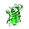
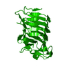
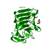
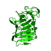
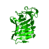
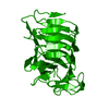
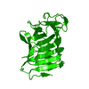
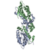
| Title | Crystal structure of truncated hemolysin A from P. mirabilis at 1.4 Angstroms resolution | ||||||
 Components Components | Hemolysin | ||||||
 Keywords Keywords | TOXIN / hemolysin / two partner secretion / beta solenoid / beta helix | ||||||
| Function / homology |  Function and homology information Function and homology informationcatalytic activity / cell outer membrane / toxin activity / killing of cells of another organism Similarity search - Function | ||||||
| Biological species |  Proteus mirabilis (bacteria) Proteus mirabilis (bacteria) | ||||||
| Method |  X-RAY DIFFRACTION / SYNCHROTRON / MOLECULAR REPLACEMENT / Resolution: 1.428 Å X-RAY DIFFRACTION / SYNCHROTRON / MOLECULAR REPLACEMENT / Resolution: 1.428 Å | ||||||
 Authors Authors | Novak, W.R.P. / Glasgow, E. / Thompson, J.R. / Weaver, T.M. | ||||||
| Funding support |  United States, 1items United States, 1items
| ||||||
 Citation Citation | Journal: Acta Crystallogr F Struct Biol Commun / Year: 2017 Title: Proteolysis of truncated hemolysin A yields a stable dimerization interface. Authors: Novak, W.R. / Bhattacharyya, B. / Grilley, D.P. / Weaver, T.M. | ||||||
| History |
|
- Structure visualization
| Structure viewer | Molecule: MolmilJmol/JSmol |
|---|
- Downloads & links
-Download
| PDBx/mmCIF format | 4w8q.cif.gz | 108.7 KB | Display | PDBx/mmCIF format |
|---|---|---|---|---|
| PDB format | pdb4w8q.ent.gz | 82.4 KB | Display | PDB format |
| PDBx/mmJSON format | 4w8q.json.gz | Tree view | PDBx/mmJSON format | |
| Others |  Other downloads Other downloads |
-Validation report
| Arichive directory | https://data.pdbj.org/pub/pdb/validation_reports/w8/4w8qftp://data.pdbj.org/pub/pdb/validation_reports/w8/4w8q | HTTPS FTP |
|---|
-Related structure data
| Related structure data | 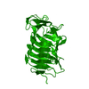 5kehC 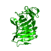 5kf3C 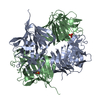 5kkdC  5sz8C  3fy3S S: Starting model for refinement C: citing same article ( |
|---|---|
| Similar structure data |


-Links
 PDBj
PDBj- Assembly
| Deposited unit | 
| |||||||||
|---|---|---|---|---|---|---|---|---|---|---|
| 1 | 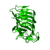
| |||||||||
| Unit cell |
| |||||||||
| Components on special symmetry positions |
|
-Components
| #1: Protein | Mass: 25479.123 Da / Num. of mol.: 1 / Fragment: UNP residues 30-265 Source method: isolated from a genetically manipulated source Source: (gene. exp.) Proteus mirabilis (bacteria) / Gene: hpmA / Plasmid: pET24a+ / Production host: |
|---|---|
| #2: Water | ChemComp-HOH /  Mass: 18.015 Da / Num. of mol.: 281 / Source method: isolated from a natural source / Formula: H2O Mass: 18.015 Da / Num. of mol.: 281 / Source method: isolated from a natural source / Formula: H2O |
| Has protein modification | Y |
-Experimental details
-Experiment
| Experiment | Method: X-RAY DIFFRACTION |
|---|
- Sample preparation
| Crystal | Density Matthews: 2.82 Å3/Da / Density % sol: 56.35 % |
|---|---|
| Crystal grow | Temperature: 298 K / Method: vapor diffusion, hanging drop / pH: 5.5 Details: 100 mM citrate pH 5.5, 100 mM NaCl, PEG 4000 (8 - 16%) |
-Data collection
| Diffraction | Mean temperature: 100 K |
|---|---|
| Diffraction source | Source: SYNCHROTRON / Site: APS / Beamline: 23-ID-B / Wavelength: 1.03 Å |
| Detector | Type: MAR scanner 300 mm plate / Detector: IMAGE PLATE / Date: Jul 3, 2014 |
| Radiation | Protocol: SINGLE WAVELENGTH / Monochromatic (M) / Laue (L): M / Scattering type: x-ray |
| Radiation wavelength | Wavelength: 1.03 Å / Relative weight: 1 |
| Reflection | Resolution: 1.428→29.7 Å / Num. obs: 49625 / % possible obs: 97.2 % / Redundancy: 6.9 % / Rmerge(I) obs: 0.078 / Net I/σ(I): 25.4 |
| Reflection shell | Resolution: 1.428→1.46 Å / Redundancy: 4.4 % / Rmerge(I) obs: 0.814 / Mean I/σ(I) obs: 2.2 / % possible all: 74.9 |
- Processing
| Software | Name: PHENIX / Version: (phenix.refine: 1.9_1692) / Classification: refinement | |||||||||||||||||||||||||||||||||||||||||||||||||||||||||||||||||||||||||||||||||||||||||||||||||||||||||
|---|---|---|---|---|---|---|---|---|---|---|---|---|---|---|---|---|---|---|---|---|---|---|---|---|---|---|---|---|---|---|---|---|---|---|---|---|---|---|---|---|---|---|---|---|---|---|---|---|---|---|---|---|---|---|---|---|---|---|---|---|---|---|---|---|---|---|---|---|---|---|---|---|---|---|---|---|---|---|---|---|---|---|---|---|---|---|---|---|---|---|---|---|---|---|---|---|---|---|---|---|---|---|---|---|---|---|
| Refinement | Method to determine structure: MOLECULAR REPLACEMENT Starting model: 3fy3 Resolution: 1.428→29.658 Å / SU ML: 0.11 / Cross valid method: FREE R-VALUE / σ(F): 0 / Phase error: 15.61 / Stereochemistry target values: ML
| |||||||||||||||||||||||||||||||||||||||||||||||||||||||||||||||||||||||||||||||||||||||||||||||||||||||||
| Solvent computation | Shrinkage radii: 0.9 Å / VDW probe radii: 1.11 Å / Solvent model: FLAT BULK SOLVENT MODEL | |||||||||||||||||||||||||||||||||||||||||||||||||||||||||||||||||||||||||||||||||||||||||||||||||||||||||
| Refinement step | Cycle: LAST / Resolution: 1.428→29.658 Å
| |||||||||||||||||||||||||||||||||||||||||||||||||||||||||||||||||||||||||||||||||||||||||||||||||||||||||
| Refine LS restraints |
| |||||||||||||||||||||||||||||||||||||||||||||||||||||||||||||||||||||||||||||||||||||||||||||||||||||||||
| LS refinement shell |
|
