- PDB-4w80: Tetrameric BAP29 vDED with disulfide bonds in crystal contacts -
+
Open data
ID or keywords:
Loading...
-
Basic information
Entry
Database: PDB / ID: 4w80
Title
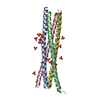
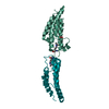
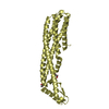
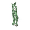
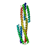
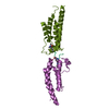
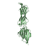
Tetrameric BAP29 vDED with disulfide bonds in crystal contacts
Components
B-cell receptor-associated protein 29
Keywords
TRANSPORT PROTEIN / Coiled coil / nanomaterial
Function / homology
Function and homology information
protein localization to endoplasmic reticulum exit site / endoplasmic reticulum to Golgi vesicle-mediated transport / intracellular protein transport / osteoblast differentiation / apoptotic process / endoplasmic reticulum membrane / membrane Similarity search - Function
B-cell receptor-associated protein 29/31 / BAP29/BAP31, transmembrane domain / Bap31/Bap29 cytoplasmic coiled-coil domain / Bap31/Bap29 transmembrane region / Bap31/Bap29 cytoplasmic coiled-coil domain / Single alpha-helices involved in coiled-coils or other helix-helix interfaces - #110 / Single alpha-helices involved in coiled-coils or other helix-helix interfaces / Up-down Bundle / Mainly Alpha Similarity search - Domain/homology
Mass: 96.063 Da / Num. of mol.: 10 / Source method: obtained synthetically / Formula: SO4
-
Experimental details
-
Experiment
Experiment
Method: X-RAY DIFFRACTION
-
Sample preparation
Crystal
Density Matthews: 4.86 Å3/Da / Density % sol: 74.67 %
Crystal grow
Temperature: 277.15 K / Method: vapor diffusion / pH: 4.6 Details: Grown in 1.8 M ammonium sulfate, 100 mM Na acetate pH 4.6 using protein purified in 20 mM Tris pH 8, 150 mM NaCl and 0.5 mM TCEP.
In the structure databanks used in Yorodumi, some data are registered as the other names, "COVID-19 virus" and "2019-nCoV". Here are the details of the virus and the list of structure data.
Jan 31, 2019. EMDB accession codes are about to change! (news from PDBe EMDB page)
EMDB accession codes are about to change! (news from PDBe EMDB page)
The allocation of 4 digits for EMDB accession codes will soon come to an end. Whilst these codes will remain in use, new EMDB accession codes will include an additional digit and will expand incrementally as the available range of codes is exhausted. The current 4-digit format prefixed with “EMD-” (i.e. EMD-XXXX) will advance to a 5-digit format (i.e. EMD-XXXXX), and so on. It is currently estimated that the 4-digit codes will be depleted around Spring 2019, at which point the 5-digit format will come into force.
The EM Navigator/Yorodumi systems omit the EMD- prefix.
Related info.:Q: What is EMD? / ID/Accession-code notation in Yorodumi/EM Navigator
Yorodumi is a browser for structure data from EMDB, PDB, SASBDB, etc.
This page is also the successor to EM Navigator detail page, and also detail information page/front-end page for Omokage search.
The word "yorodu" (or yorozu) is an old Japanese word meaning "ten thousand". "mi" (miru) is to see.
Related info.:EMDB / PDB / SASBDB / Comparison of 3 databanks / Yorodumi Search / Aug 31, 2016. New EM Navigator & Yorodumi / Yorodumi Papers / Jmol/JSmol / Function and homology information / Changes in new EM Navigator and Yorodumi
 Movie
Movie Controller


 Open data
 Basic information
 Components
 Keywords
 Function and homology information
 Homo sapiens (human)
 X-RAY DIFFRACTION /
  Authors
 Citation
 Structure visualization
 Downloads & links
 Other downloads


 PDBj
PDBj Assembly


 Mass: 96.063 Da / Num. of mol.: 10 / Source method: obtained synthetically / Formula: SO4
 Sample preparation
 / Beamline: I04-1 / Wavelength: 0.92 Å
 Processing