Component-ID: 1 / Ens-ID: 1 / End auth comp-ID: ALA / End label comp-ID: ALA
Dom-ID
Beg auth comp-ID
Beg label comp-ID
Selection details
Auth asym-ID
Label asym-ID
Auth seq-ID
Label seq-ID
1
GLY
GLY
chainA
A
B
40 - 408
40 - 408
2
ASN
ASN
chainB
B
A
39 - 408
39 - 408
-
Components
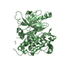
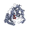
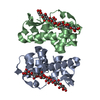
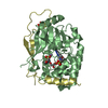
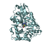
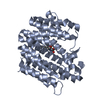
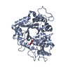
#1: Protein
Chitinase
Mass: 43334.617 Da / Num. of mol.: 2 Source method: isolated from a genetically manipulated source Details: Secretion signal peptide has been recognized and processed by E. coli. Source: (gene. exp.) Streptomyces thermoviolaceus (bacteria) Gene: chi40 / Production host: Escherichia coli (E. coli) / References: UniProt: Q56077, chitinase
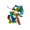
Method to determine structure: MOLECULAR REPLACEMENT Starting model: The model structure was generated by the GeneSilico Metaserver. Resolution: 2.771→49 Å / SU ML: 0.31 / Cross valid method: FREE R-VALUE / σ(F): 1.36 / Phase error: 22.69 / Stereochemistry target values: ML
Rfactor
Num. reflection
% reflection
Selection details
Rfree
0.2084
995
3 %
Random selection
Rwork
0.1498
32149
-
-
obs
0.1515
33144
99.4 %
-
Solvent computation
Shrinkage radii: 0.9 Å / VDW probe radii: 1.11 Å / Solvent model: FLAT BULK SOLVENT MODEL
In the structure databanks used in Yorodumi, some data are registered as the other names, "COVID-19 virus" and "2019-nCoV". Here are the details of the virus and the list of structure data.
Jan 31, 2019. EMDB accession codes are about to change! (news from PDBe EMDB page)
EMDB accession codes are about to change! (news from PDBe EMDB page)
The allocation of 4 digits for EMDB accession codes will soon come to an end. Whilst these codes will remain in use, new EMDB accession codes will include an additional digit and will expand incrementally as the available range of codes is exhausted. The current 4-digit format prefixed with “EMD-” (i.e. EMD-XXXX) will advance to a 5-digit format (i.e. EMD-XXXXX), and so on. It is currently estimated that the 4-digit codes will be depleted around Spring 2019, at which point the 5-digit format will come into force.
The EM Navigator/Yorodumi systems omit the EMD- prefix.
Related info.:Q: What is EMD? / ID/Accession-code notation in Yorodumi/EM Navigator
Yorodumi is a browser for structure data from EMDB, PDB, SASBDB, etc.
This page is also the successor to EM Navigator detail page, and also detail information page/front-end page for Omokage search.
The word "yorodu" (or yorozu) is an old Japanese word meaning "ten thousand". "mi" (miru) is to see.
Related info.:EMDB / PDB / SASBDB / Comparison of 3 databanks / Yorodumi Search / Aug 31, 2016. New EM Navigator & Yorodumi / Yorodumi Papers / Jmol/JSmol / Function and homology information / Changes in new EM Navigator and Yorodumi
 Movie
Movie Controller

 Yorodumi
 Open data
 Basic information
 Components
 Keywords
 Function and homology information
 Streptomyces thermoviolaceus (bacteria)
 X-RAY DIFFRACTION /
  Authors
 Poland, 1items
  Citation
 Structure visualization
 Downloads & links
 Other downloads
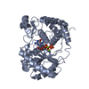


 PDBj
PDBj Assembly


 Mass: 102.046 Da / Num. of mol.: 2 / Source method: obtained synthetically / Formula: C3H2O4
 Mass: 18.015 Da / Num. of mol.: 74 / Source method: isolated from a natural source / Formula: H2O
 Sample preparation
 / Beamline: I911-3 / Wavelength: 1 Å
 Processing