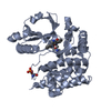
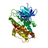
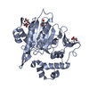
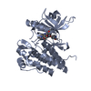
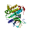
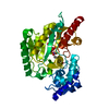
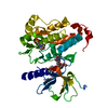
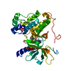
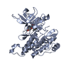
登録情報 データベース : PDB / ID : 3lcdタイトル Inhibitor Bound to A DFG-In structure of the Kinase Domain of CSF-1R Macrophage colony-stimulating factor 1 receptor キーワード / / / / / / / / / / / / / 機能・相同性 分子機能 ドメイン・相同性 構成要素
/ / / / / / / / / / / / / / / / / / / / / / / / / / / / / / / / / / / / / / / / / / / / / / / / / / / / / / / / / / / / / / / / / / / / / / / / / / / / / / / / / / / / / / / / / / / / / / / / / / / / / / / / / / / / / 生物種 Homo sapiens (ヒト)手法 / / / 解像度 : 2.5 Å データ登録者 Kamtekar, S. / Day, J.E. / Reitz, B.A. / Mathis, K.J. / Meyers, M.J. ジャーナル : Bioorg.Med.Chem.Lett. / 年 : 2010タイトル : Structure-based drug design enables conversion of a DFG-in binding CSF-1R kinase inhibitor to a DFG-out binding mode著者: Meyers, M.J. / Pelc, M. / Kamtekar, S. / Day, J. / Poda, G.I. / Hall, M.K. / Michener, M.L. / Reitz, B.A. / Mathis, K.J. / Pierce, B.S. / Parikh, M.D. / Mischke, D.A. / Long, S.A. / Parlow, J. ... 著者 : Meyers, M.J. / Pelc, M. / Kamtekar, S. / Day, J. / Poda, G.I. / Hall, M.K. / Michener, M.L. / Reitz, B.A. / Mathis, K.J. / Pierce, B.S. / Parikh, M.D. / Mischke, D.A. / Long, S.A. / Parlow, J.J. / Anderson, D.R. / Thorarensen, A. 履歴 登録 2010年1月10日 登録サイト / 処理サイト 改定 1.0 2010年3月2日 Provider / タイプ 改定 1.1 2011年7月13日 Group / Version format compliance改定 1.2 2017年7月26日 Group / Refinement description / Source and taxonomyカテゴリ / entity_src_gen / software / Item 改定 1.3 2024年10月16日 Group Data collection / Database references ... Data collection / Database references / Derived calculations / Structure summary カテゴリ chem_comp_atom / chem_comp_bond ... chem_comp_atom / chem_comp_bond / database_2 / pdbx_entry_details / struct_conn / struct_ref_seq_dif / struct_site Item _database_2.pdbx_DOI / _database_2.pdbx_database_accession ... _database_2.pdbx_DOI / _database_2.pdbx_database_accession / _struct_conn.pdbx_leaving_atom_flag / _struct_ref_seq_dif.details / _struct_site.pdbx_auth_asym_id / _struct_site.pdbx_auth_comp_id / _struct_site.pdbx_auth_seq_id
すべて表示 表示を減らす
 ムービー
ムービー コントローラー


 データを開く
 基本情報
基本情報 要素
要素 キーワード
キーワード 機能・相同性情報
 Homo sapiens (ヒト)
 X線回折 /
  データ登録者
 引用
引用 構造の表示
構造の表示 ダウンロードとリンク
 その他のダウンロード

 PDBj
PDBj


 集合体
集合体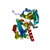

 Spodoptera frugiperda (ツマジロクサヨトウ)
 分子量: 96.063 Da / 分子数: 2 / 由来タイプ: 合成 / 式: SO4
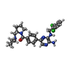
 分子量: 525.473 Da / 分子数: 1 / 由来タイプ: 合成 / 式: C27H30Cl2N6O
 分子量: 18.015 Da / 分子数: 44 / 由来タイプ: 天然 / 式: H2O
 試料調製
試料調製 / ビームライン: 21-ID-F / 波長: 1.07804 Å
 解析
解析