 Movie
Movie Controller

[English] 日本語
 Yorodumi
- PDB-4v8w: Structure and conformational variability of the Mycobacterium tub... -
+ Open data

- Basic information
| Entry | Database: PDB / ID: 4v8w | ||||||||||||
|---|---|---|---|---|---|---|---|---|---|---|---|---|---|
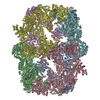
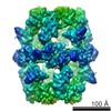
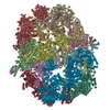
| Title | Structure and conformational variability of the Mycobacterium tuberculosis fatty acid synthase multienzyme complex | ||||||||||||
 Components Components | TYPE-I FATTY ACID SYNTHASE | ||||||||||||
 Keywords Keywords | HYDROLASE / FATTY ACID SYNTHESIS / SAMPLE HETEROGENEITY / PROTEIN FLEXIBILITY / CODIMENSIONAL PRINCIPAL COMPONENT ANALYSIS | ||||||||||||
| Function / homology | FLAVIN MONONUCLEOTIDE Function and homology information Function and homology information | ||||||||||||
| Biological species |   MYCOBACTERIUM TUBERCULOSIS (bacteria) MYCOBACTERIUM TUBERCULOSIS (bacteria) | ||||||||||||
| Method | ELECTRON MICROSCOPY / single particle reconstruction / cryo EM / Resolution: 17.5 Å | ||||||||||||
 Authors Authors | Ciccarelli, L. / Connell, S.R. / Enderle, M. / Mills, D.J. / Vonck, J. / Grininger, M. | ||||||||||||
 Citation Citation | Journal: Structure / Year: 2013 Title: Structure and conformational variability of the mycobacterium tuberculosis fatty acid synthase multienzyme complex. Authors: Luciano Ciccarelli / Sean R Connell / Mathias Enderle / Deryck J Mills / Janet Vonck / Martin Grininger /  Abstract: Antibiotic therapy in response to Mycobacterium tuberculosis infections targets de novo fatty acid biosynthesis, which is orchestrated by a 1.9 MDa type I fatty acid synthase (FAS). Here, we ...Antibiotic therapy in response to Mycobacterium tuberculosis infections targets de novo fatty acid biosynthesis, which is orchestrated by a 1.9 MDa type I fatty acid synthase (FAS). Here, we characterize M. tuberculosis FAS by single-particle cryo-electron microscopy and interpret the data by docking the molecular models of yeast and Mycobacterium smegmatis FAS. Our analysis reveals a porous barrel-like structure of considerable conformational variability that is illustrated by the identification of several conformational states with altered topology in the multienzymatic assembly. This demonstrates that the barrel-like structure of M. tuberculosis FAS is not just a static scaffold for the catalytic domains, but may play an active role in coordinating fatty acid synthesis. The conception of M. tuberculosis FAS as a highly dynamic assembly of domains revises the view on bacterial type I fatty acid synthesis and might inspire new strategies for inhibition of de novo fatty acid synthesis in M. tuberculosis. | ||||||||||||
| History |
| ||||||||||||
| Remark 700 | SHEET DETERMINATION METHOD: DSSP THE SHEETS PRESENTED AS "DB" IN EACH CHAIN ON SHEET RECORDS BELOW ... SHEET DETERMINATION METHOD: DSSP THE SHEETS PRESENTED AS "DB" IN EACH CHAIN ON SHEET RECORDS BELOW IS ACTUALLY AN 8-STRANDED BARREL THIS IS REPRESENTED BY A 9-STRANDED SHEET IN WHICH THE FIRST AND LAST STRANDS ARE IDENTICAL. THE SHEETS PRESENTED AS "ED" IN EACH CHAIN ON SHEET RECORDS BELOW IS ACTUALLY AN 8-STRANDED BARREL THIS IS REPRESENTED BY A 9-STRANDED SHEET IN WHICH THE FIRST AND LAST STRANDS ARE IDENTICAL. THE SHEETS PRESENTED AS "FD" IN EACH CHAIN ON SHEET RECORDS BELOW IS ACTUALLY AN 8-STRANDED BARREL THIS IS REPRESENTED BY A 9-STRANDED SHEET IN WHICH THE FIRST AND LAST STRANDS ARE IDENTICAL. |
- Structure visualization
| Movie |
Movie viewer |
|---|---|
| Structure viewer | Molecule: MolmilJmol/JSmol |
- Downloads & links
-Download
| PDBx/mmCIF format | 4v8w.cif.gz | 2.6 MB | Display | PDBx/mmCIF format |
|---|---|---|---|---|
| PDB format | pdb4v8w.ent.gz | Display | PDB format | |
| PDBx/mmJSON format | 4v8w.json.gz | Tree view | PDBx/mmJSON format | |
| Others |  Other downloads Other downloads |
-Validation report
| Arichive directory | https://data.pdbj.org/pub/pdb/validation_reports/v8/4v8wftp://data.pdbj.org/pub/pdb/validation_reports/v8/4v8w | HTTPS FTP |
|---|
-Related structure data
| Related structure data | 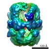 2357MC 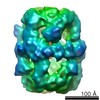 2358C 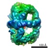 2359C  4v8vC C: citing same article ( M: map data used to model this data |
|---|---|
| Similar structure data |


-Links
 PDBj
PDBj- Assembly
| Deposited unit | 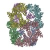
|
|---|---|
| 1 |
|
-Components
| #1: Protein | Mass: 329887.969 Da / Num. of mol.: 6 / Source method: isolated from a natural source / Source: (natural) MYCOBACTERIUM TUBERCULOSIS (bacteria)#2: Chemical | ChemComp-FMN /   Mass: 456.344 Da / Num. of mol.: 6 / Source method: obtained synthetically / Formula: C17H21N4O9P Mass: 456.344 Da / Num. of mol.: 6 / Source method: obtained synthetically / Formula: C17H21N4O9PSequence details | UNP ACCESSION A0R1H7 (M. SMEGMATIS) IS MODELLED TO FIT THE M. TUBERCULOS | |
|---|
-Experimental details
-Experiment
| Experiment | Method: ELECTRON MICROSCOPY |
|---|---|
| EM experiment | Aggregation state: PARTICLE / 3D reconstruction method: single particle reconstruction |
- Sample preparation
| Component | Name: Mycobacterium tuberculosis fatty acid synthase multienzyme complex Type: COMPLEX |
|---|---|
| Specimen | Conc.: 1 mg/ml / Embedding applied: NO / Shadowing applied: NO / Staining applied: NO / Vitrification applied: YES |
| Specimen support | Details: OTHER |
| Vitrification | Instrument: FEI VITROBOT MARK III / Cryogen name: ETHANE |
- Electron microscopy imaging
| Experimental equipment |  Model: Tecnai Polara / Image courtesy: FEI Company |
|---|---|
| Microscopy | Model: FEI POLARA 300 / Date: Dec 16, 2010 |
| Electron gun | Electron source:  FIELD EMISSION GUN / Accelerating voltage: 200 kV / Illumination mode: FLOOD BEAM FIELD EMISSION GUN / Accelerating voltage: 200 kV / Illumination mode: FLOOD BEAM |
| Electron lens | Mode: BRIGHT FIELD / Nominal magnification: 59000 X / Nominal defocus max: 4500 nm / Nominal defocus min: 1800 nm |
| Image recording | Film or detector model: KODAK SO-163 FILM |
| Radiation wavelength | Relative weight: 1 |
- Processing
| EM software |
| ||||||||||||
|---|---|---|---|---|---|---|---|---|---|---|---|---|---|
| Symmetry | Point symmetry: C1 (asymmetric) | ||||||||||||
| 3D reconstruction | Resolution: 17.5 Å / Num. of particles: 9136 Details: THE MODEL IS DERIVED FROM PDB ENTRY 3ZEN BOEHRINGER ET AL., 2013 AND REPRESENTS A RIGID BODY DOCKING OF THE MODEL INTO A CRYO-EM MAP. IN CHAIN F THE MAT AND DH DOMAINS WHERE FIT ...Details: THE MODEL IS DERIVED FROM PDB ENTRY 3ZEN BOEHRINGER ET AL., 2013 AND REPRESENTS A RIGID BODY DOCKING OF THE MODEL INTO A CRYO-EM MAP. IN CHAIN F THE MAT AND DH DOMAINS WHERE FIT INDEPENDENTLY OF THE REST OF THE MODEL. IN CHAIN D, RESIDUES 1- 400 (AT DOMAIN) WERE DELETED AS THE DENSITY OF THE EM MAP IN THAT REGION IS FRAGMENTED DUE TO FLEXIBILITY. SUBMISSION BASED ON EXPERIMENTAL DATA FROM EMDB EMD-2357. (DEPOSITION ID: 11618). Symmetry type: POINT | ||||||||||||
| Atomic model building | Protocol: RIGID BODY FIT / Space: REAL / Details: METHOD--RIGID BODY REFINEMENT PROTOCOL--RIGID BODY | ||||||||||||
| Atomic model building | PDB-ID: 3ZEN 3zen Accession code: 3ZEN / Source name: PDB / Type: experimental model | ||||||||||||
| Refinement | Highest resolution: 17.5 Å | ||||||||||||
| Refinement step | Cycle: LAST / Highest resolution: 17.5 Å
|
