[English] 日本語
Yorodumi
- PDB-4v7d: Structure of the Ribosome with Elongation Factor G Trapped in the... -

+
Open data


ID or keywords:

Loading...

-
Basic information

Entry
Database: PDB / ID: 4v7d
TitleStructure of the Ribosome with Elongation Factor G Trapped in the Pre-Translocation State (pre-translocation 70S*tRNA*EF-G structure)
Components
  • (30S ribosomal protein ...) x 20
  • (50S ribosomal protein ...) x 32
  • 16S ribosomal RNA
  • 23S ribosomal RNA
  • 5S ribosomal RNA
  • Elongation Factor G
  • mRNA
  • tRNA
  • viomycin
KeywordsTRANSLATION/antibiotic / translation / EF-G / single particle analysis / pre-translocation translation complex / viomycin / TRANSLATION-antibiotic complex
Function / homology
Function and homology information


ribosome disassembly / translational elongation / guanosine tetraphosphate binding / stringent response / mRNA base-pairing translational repressor activity / ornithine decarboxylase inhibitor activity / transcription antitermination factor activity, RNA binding / misfolded RNA binding / Group I intron splicing / RNA folding ...ribosome disassembly / translational elongation / guanosine tetraphosphate binding / stringent response / mRNA base-pairing translational repressor activity / ornithine decarboxylase inhibitor activity / transcription antitermination factor activity, RNA binding / misfolded RNA binding / Group I intron splicing / RNA folding / transcriptional attenuation / endoribonuclease inhibitor activity / RNA-binding transcription regulator activity / positive regulation of ribosome biogenesis / negative regulation of cytoplasmic translation / translation elongation factor activity / translational termination / DnaA-L2 complex / four-way junction DNA binding / translation repressor activity / negative regulation of translational initiation / negative regulation of DNA-templated DNA replication initiation / regulation of mRNA stability / ribosome assembly / mRNA regulatory element binding translation repressor activity / positive regulation of RNA splicing / assembly of large subunit precursor of preribosome / DNA endonuclease activity / transcription elongation factor complex / cytosolic ribosome assembly / regulation of DNA-templated transcription elongation / transcription antitermination / response to reactive oxygen species / regulation of cell growth / DNA-templated transcription termination / maintenance of translational fidelity / Hydrolases; Acting on acid anhydrides; Acting on GTP to facilitate cellular and subcellular movement / response to radiation / ribosomal large subunit assembly / mRNA 5'-UTR binding / large ribosomal subunit / ribosome biogenesis / ribosome binding / regulation of translation / ribosomal small subunit biogenesis / ribosomal small subunit assembly / small ribosomal subunit / small ribosomal subunit rRNA binding / transferase activity / 5S rRNA binding / large ribosomal subunit rRNA binding / cytosolic small ribosomal subunit / cytosolic large ribosomal subunit / tRNA binding / cytoplasmic translation / molecular adaptor activity / rRNA binding / negative regulation of translation / ribosome / structural constituent of ribosome / translation / response to antibiotic / GTPase activity / negative regulation of DNA-templated transcription / mRNA binding / GTP binding / protein homodimerization activity / DNA binding / RNA binding / zinc ion binding / membrane / cytoplasm / cytosol
Similarity search - Function
: / Translation elongation factor EFG/EF2 / Elongation factor G, domain III / EFG, domain V / Ribosomal protein L7/L12, oligomerisation / Ribosomal protein L7/L12, oligomerisation domain superfamily / Ribosomal protein L7/L12 dimerisation domain / Ribosomal protein L7/L12 / Ribosomal protein L7/L12, C-terminal / Ribosomal protein L7/L12 C-terminal domain ...: / Translation elongation factor EFG/EF2 / Elongation factor G, domain III / EFG, domain V / Ribosomal protein L7/L12, oligomerisation / Ribosomal protein L7/L12, oligomerisation domain superfamily / Ribosomal protein L7/L12 dimerisation domain / Ribosomal protein L7/L12 / Ribosomal protein L7/L12, C-terminal / Ribosomal protein L7/L12 C-terminal domain / Ribosomal protein L7/L12, C-terminal/adaptor protein ClpS-like / Elongation Factor G, domain II / Elongation Factor G, domain III / Ribosomal protein L1, bacterial-type / Translation elongation factor EFG/EF2, domain IV / Elongation factor G, domain IV / Elongation factor G, domain IV / Elongation factor G C-terminus / Ribosomal protein L10, eubacterial, conserved site / Ribosomal protein L10 signature. / Elongation factor EFG, domain V-like / Elongation factor G C-terminus / Ribosomal protein L10 / EF-G domain III/V-like / Tr-type G domain, conserved site / Translational (tr)-type guanine nucleotide-binding (G) domain signature. / : / Ribosomal protein L1, conserved site / Ribosomal protein L1 signature. / Ribosomal protein L1 / Ribosomal protein L1, 3-layer alpha/beta-sandwich / Translation elongation factor EFTu-like, domain 2 / Ribosomal protein S21, conserved site / Ribosomal protein S21 signature. / Ribosomal protein L25, short-form / Ribosomal protein S14, bacterial/plastid / Elongation factor Tu domain 2 / Ribosomal protein L1-like / Ribosomal protein L1/ribosomal biogenesis protein / Ribosomal protein L1p/L10e family / Ribosomal protein L11, bacterial-type / Ribosomal protein S21 superfamily / Ribosomal protein S21 / Ribosomal protein S16, conserved site / Ribosomal protein S16 signature. / Translational (tr)-type GTP-binding domain / Elongation factor Tu GTP binding domain / Translational (tr)-type guanine nucleotide-binding (G) domain profile. / Ribosomal protein S21 / Ribosomal protein L11, conserved site / Ribosomal protein L11 signature. / Ribosomal protein L21, conserved site / Ribosomal protein L21 signature. / Ribosomal protein L10-like domain superfamily / Ribosomal protein L10 / Ribosomal protein L16 signature 1. / Ribosomal protein L10P / : / Ribosomal protein L6, conserved site / Ribosomal protein L6 signature 1. / Ribosomal protein L16, conserved site / Ribosomal protein L16 signature 2. / Ribosomal protein L9 signature. / Ribosomal protein L9, bacteria/chloroplast / Ribosomal protein L9, C-terminal / Ribosomal protein L9, C-terminal domain / Ribosomal protein L17 signature. / Ribosomal protein L9, C-terminal domain superfamily / Ribosomal L25p family / Ribosomal protein L25 / Ribosomal protein L11, N-terminal / Ribosomal protein L11, N-terminal domain / Ribosomal protein L11/L12 / Ribosomal protein L11, C-terminal / Ribosomal protein L11, C-terminal domain superfamily / Ribosomal protein L11/L12, N-terminal domain superfamily / Ribosomal protein L11, RNA binding domain / Ribosomal protein L11/L12 / Ribosomal protein L36 signature. / Ribosomal protein L28/L24 superfamily / Ribosomal protein L25/Gln-tRNA synthetase, N-terminal / Ribosomal protein L25/Gln-tRNA synthetase, anti-codon-binding domain superfamily / Ribosomal protein L32p, bacterial type / Ribosomal protein L9, N-terminal domain superfamily / Ribosomal protein L9 / Ribosomal protein L9, N-terminal / Ribosomal protein L9, N-terminal domain / Ribosomal protein L28 / Ribosomal protein L35, conserved site / Ribosomal protein L35 signature. / Ribosomal protein L33, conserved site / Ribosomal protein L33 signature. / Ribosomal protein L35, non-mitochondrial / Ribosomal protein L5, bacterial-type / Ribosomal protein L6, bacterial-type / Ribosomal protein L18, bacterial-type / Ribosomal protein L19, conserved site / Ribosomal protein L19 signature. / Ribosomal protein L36 / Ribosomal protein L36 superfamily
Similarity search - Domain/homology
Viomycin / : / : / : / : / : / RNA / RNA (> 10) / RNA (> 100) / RNA (> 1000) ...Viomycin / : / : / : / : / : / RNA / RNA (> 10) / RNA (> 100) / RNA (> 1000) / Small ribosomal subunit protein bS6 / Small ribosomal subunit protein uS7 / Large ribosomal subunit protein uL15 / Elongation factor G / Large ribosomal subunit protein uL10 / Large ribosomal subunit protein uL11 / Large ribosomal subunit protein bL12 / Large ribosomal subunit protein bL19 / Large ribosomal subunit protein uL1 / Large ribosomal subunit protein bL20 / Large ribosomal subunit protein bL27 / Large ribosomal subunit protein bL28 / Large ribosomal subunit protein uL29 / Large ribosomal subunit protein bL32 / Large ribosomal subunit protein bL33 / Large ribosomal subunit protein bL34 / Large ribosomal subunit protein bL35 / Large ribosomal subunit protein bL36A / Large ribosomal subunit protein bL9 / Small ribosomal subunit protein uS10 / Small ribosomal subunit protein uS11 / Small ribosomal subunit protein uS12 / Small ribosomal subunit protein uS13 / Small ribosomal subunit protein bS16 / Small ribosomal subunit protein bS18 / Small ribosomal subunit protein uS19 / Small ribosomal subunit protein bS20 / Small ribosomal subunit protein uS2 / Small ribosomal subunit protein uS3 / Small ribosomal subunit protein uS4 / Small ribosomal subunit protein uS5 / Small ribosomal subunit protein uS8 / Small ribosomal subunit protein uS9 / Large ribosomal subunit protein uL13 / Large ribosomal subunit protein uL14 / Large ribosomal subunit protein uL16 / Large ribosomal subunit protein uL23 / Small ribosomal subunit protein uS15 / Large ribosomal subunit protein bL17 / Large ribosomal subunit protein bL21 / Large ribosomal subunit protein uL30 / Large ribosomal subunit protein uL6 / Small ribosomal subunit protein uS14 / Small ribosomal subunit protein uS17 / Large ribosomal subunit protein uL18 / Large ribosomal subunit protein uL2 / Large ribosomal subunit protein uL3 / Large ribosomal subunit protein uL24 / Large ribosomal subunit protein uL4 / Large ribosomal subunit protein uL22 / Large ribosomal subunit protein uL5 / Small ribosomal subunit protein bS21 / Large ribosomal subunit protein bL25
Similarity search - Component
Biological speciesEscherichia coli (E. coli)
Synthetic (others)
Streptomyces (bacteria)
MethodELECTRON MICROSCOPY / single particle reconstruction / cryo EM / Resolution: 7.6 Å
AuthorsBrilot, A.F. / Korostelev, A.A. / Ermolenko, D.N. / Grigorieff, N.
CitationJournal: Proc Natl Acad Sci U S A / Year: 2013
Title: Structure of the ribosome with elongation factor G trapped in the pretranslocation state.
Authors: Axel F Brilot / Andrei A Korostelev / Dmitri N Ermolenko / Nikolaus Grigorieff /
Abstract: During protein synthesis, tRNAs and their associated mRNA codons move sequentially on the ribosome from the A (aminoacyl) site to the P (peptidyl) site to the E (exit) site in a process catalyzed by ...During protein synthesis, tRNAs and their associated mRNA codons move sequentially on the ribosome from the A (aminoacyl) site to the P (peptidyl) site to the E (exit) site in a process catalyzed by a universally conserved ribosome-dependent GTPase [elongation factor G (EF-G) in prokaryotes and elongation factor 2 (EF-2) in eukaryotes]. Although the high-resolution structure of EF-G bound to the posttranslocation ribosome has been determined, the pretranslocation conformation of the ribosome bound with EF-G and A-site tRNA has evaded visualization owing to the transient nature of this state. Here we use electron cryomicroscopy to determine the structure of the 70S ribosome with EF-G, which is trapped in the pretranslocation state using antibiotic viomycin. Comparison with the posttranslocation ribosome shows that the small subunit of the pretranslocation ribosome is rotated by ∼12° relative to the large subunit. Domain IV of EF-G is positioned in the cleft between the body and head of the small subunit outwardly of the A site and contacts the A-site tRNA. Our findings suggest a model in which domain IV of EF-G promotes the translocation of tRNA from the A to the P site as the small ribosome subunit spontaneously rotates back from the hybrid, rotated state into the nonrotated posttranslocation state.
History
DepositionNov 21, 2013Deposition site: RCSB / Processing site: RCSB
Revision 1.0Jul 9, 2014Provider: repository / Type: Initial release
SupersessionDec 10, 2014ID: 3J5W, 3J5X
Revision 1.1Dec 10, 2014Group: Other
Revision 1.2Dec 17, 2014Group: Source and taxonomy / Structure summary
Revision 1.3Apr 15, 2015Group: Other
Revision 1.4Jul 18, 2018Group: Data collection / Category: em_software / Item: _em_software.image_processing_id / _em_software.name
Revision 1.5Dec 18, 2019Group: Database references / Derived calculations / Other / Category: atom_sites / struct_conn / struct_ref_seq_dif
Item: _atom_sites.fract_transf_matrix[1][1] / _atom_sites.fract_transf_matrix[2][2] ..._atom_sites.fract_transf_matrix[1][1] / _atom_sites.fract_transf_matrix[2][2] / _atom_sites.fract_transf_matrix[3][3] / _struct_conn.pdbx_leaving_atom_flag / _struct_ref_seq_dif.details

-
Structure visualization

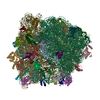
Movie
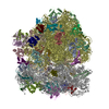
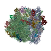
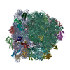
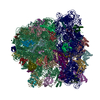
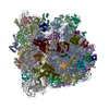
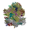
  • Deposited structure unit
  • Imaged by Jmol
  • Download
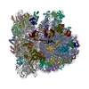
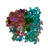
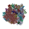
  • Superimposition on EM map
  • EMDB-5800
  • Imaged by UCSF Chimera
  • Download
Movie viewer
Structure viewerMolecule:
MolmilJmol/JSmol

Downloads & links

-
Assembly

Deposited unit
AA: 23S ribosomal RNA
AB: 5S ribosomal RNA
AC: 50S ribosomal protein L1
AD: 50S ribosomal protein L2
AE: 50S ribosomal protein L3
AF: 50S ribosomal protein L4
AG: 50S ribosomal protein L5
AH: 50S ribosomal protein L6
AI: 50S ribosomal protein L9
AJ: 50S ribosomal protein L10
AK: 50S ribosomal protein L11
AL: 50S ribosomal protein L12
AM: 50S ribosomal protein L13
AN: 50S ribosomal protein L14
AO: 50S ribosomal protein L15
AP: 50S ribosomal protein L16
AQ: 50S ribosomal protein L17
AR: 50S ribosomal protein L18
AS: 50S ribosomal protein L19
AT: 50S ribosomal protein L20
AU: 50S ribosomal protein L21
AV: 50S ribosomal protein L22
AW: 50S ribosomal protein L23
AX: 50S ribosomal protein L24
AY: 50S ribosomal protein L25
AZ: 50S ribosomal protein L27
A1: 50S ribosomal protein L28
A2: 50S ribosomal protein L29
A3: 50S ribosomal protein L30
A4: 50S ribosomal protein L32
A5: 50S ribosomal protein L33
A6: 50S ribosomal protein L34
A7: 50S ribosomal protein L35
A8: 50S ribosomal protein L36
BA: 16S ribosomal RNA
BB: 30S ribosomal protein S2
BC: 30S ribosomal protein S3
BD: 30S ribosomal protein S4
BE: 30S ribosomal protein S5
BF: 30S ribosomal protein S6
BG: 30S ribosomal protein S7
BH: 30S ribosomal protein S8
BI: 30S ribosomal protein S9
BJ: 30S ribosomal protein S10
BK: 30S ribosomal protein S11
BL: 30S ribosomal protein S12
BM: 30S ribosomal protein S13
BN: 30S ribosomal protein S14
BO: 30S ribosomal protein S15
BP: 30S ribosomal protein S16
BQ: 30S ribosomal protein S17
BR: 30S ribosomal protein S18
BS: 30S ribosomal protein S19
BT: 30S ribosomal protein S20
BU: 30S ribosomal protein S21
BV: tRNA
BW: tRNA
BX: mRNA
BZ: Elongation Factor G
BY: viomycin
hetero molecules


Theoretical massNumber of molelcules
Total (without water)2,330,68761
Polymers2,330,62260
Non-polymers651
Water00
1


  • Idetical with deposited unit
  • defined by author
TypeNameSymmetry operationNumber
identity operation1_555x,y,z1

-
Components

-
RNA chain , 5 types, 6 molecules AAABBABVBWBX

#1: RNA chain 23S ribosomal RNA


Mass: 941306.188 Da / Num. of mol.: 1 / Source method: isolated from a natural source / Source: (natural) Escherichia coli (E. coli) / Strain: MRE600 / References: GenBank: 33357927
#2: RNA chain 5S ribosomal RNA


Mass: 38483.926 Da / Num. of mol.: 1 / Source method: isolated from a natural source / Source: (natural) Escherichia coli (E. coli) / Strain: MRE600 / References: GenBank: CP000948.1
#35: RNA chain 16S ribosomal RNA


Mass: 499690.031 Da / Num. of mol.: 1 / Source method: isolated from a natural source / Source: (natural) Escherichia coli (E. coli) / Strain: MRE600 / References: GenBank: J01695.2
#56: RNA chain tRNA


Mass: 24485.539 Da / Num. of mol.: 2 / Fragment: SEE REMARK 999 / Source method: isolated from a natural source / Source: (natural) Escherichia coli (E. coli) / Strain: K-12 / References: GenBank: AP009048.1
#57: RNA chain mRNA


Mass: 5781.499 Da / Num. of mol.: 1 / Fragment: SEE REMARK 999 / Source method: obtained synthetically / Source: (synth.) Synthetic (others)

+
50S ribosomal protein ... , 32 types, 32 molecules ACADAEAFAGAHAIAJAKALAMANAOAPAQARASATAUAVAWAXAYAZA1A2A3A4A5A6A7A8

#3: Protein 50S ribosomal protein L1


Mass: 24634.465 Da / Num. of mol.: 1 / Source method: isolated from a natural source / Source: (natural) Escherichia coli (E. coli) / Strain: MRE600 / References: UniProt: P0A7L0
#4: Protein 50S ribosomal protein L2


Mass: 29792.424 Da / Num. of mol.: 1 / Source method: isolated from a natural source / Source: (natural) Escherichia coli (E. coli) / Strain: MRE600 / References: UniProt: P60422
#5: Protein 50S ribosomal protein L3


Mass: 22277.535 Da / Num. of mol.: 1 / Source method: isolated from a natural source / Source: (natural) Escherichia coli (E. coli) / Strain: MRE600 / References: UniProt: P60438
#6: Protein 50S ribosomal protein L4


Mass: 22121.566 Da / Num. of mol.: 1 / Source method: isolated from a natural source / Source: (natural) Escherichia coli (E. coli) / Strain: MRE600 / References: UniProt: P60723
#7: Protein 50S ribosomal protein L5


Mass: 20202.416 Da / Num. of mol.: 1 / Source method: isolated from a natural source / Source: (natural) Escherichia coli (E. coli) / Strain: MRE600 / References: UniProt: P62399
#8: Protein 50S ribosomal protein L6


Mass: 18801.598 Da / Num. of mol.: 1 / Source method: isolated from a natural source / Source: (natural) Escherichia coli (E. coli) / Strain: MRE600 / References: UniProt: P0AG55
#9: Protein 50S ribosomal protein L9


Mass: 15789.020 Da / Num. of mol.: 1 / Source method: isolated from a natural source / Source: (natural) Escherichia coli (E. coli) / Strain: MRE600 / References: UniProt: P0A7R1
#10: Protein 50S ribosomal protein L10


Mass: 17736.596 Da / Num. of mol.: 1 / Source method: isolated from a natural source / Source: (natural) Escherichia coli (E. coli) / Strain: MRE600 / References: UniProt: P0A7J3
#11: Protein 50S ribosomal protein L11


Mass: 14763.165 Da / Num. of mol.: 1 / Source method: isolated from a natural source / Source: (natural) Escherichia coli (E. coli) / Strain: MRE600 / References: UniProt: P0A7J7
#12: Protein 50S ribosomal protein L12


Mass: 12177.960 Da / Num. of mol.: 1 / Source method: isolated from a natural source / Source: (natural) Escherichia coli (E. coli) / Strain: MRE600 / References: UniProt: P0A7K2
#13: Protein 50S ribosomal protein L13


Mass: 16050.606 Da / Num. of mol.: 1 / Source method: isolated from a natural source / Source: (natural) Escherichia coli (E. coli) / Strain: MRE600 / References: UniProt: P0AA10
#14: Protein 50S ribosomal protein L14


Mass: 13565.067 Da / Num. of mol.: 1 / Source method: isolated from a natural source / Source: (natural) Escherichia coli (E. coli) / Strain: MRE600 / References: UniProt: P0ADY3
#15: Protein 50S ribosomal protein L15


Mass: 15008.471 Da / Num. of mol.: 1 / Source method: isolated from a natural source / Source: (natural) Escherichia coli (E. coli) / Strain: MRE600 / References: UniProt: P02413
#16: Protein 50S ribosomal protein L16


Mass: 15312.269 Da / Num. of mol.: 1 / Source method: isolated from a natural source / Source: (natural) Escherichia coli (E. coli) / Strain: MRE600 / References: UniProt: P0ADY7
#17: Protein 50S ribosomal protein L17


Mass: 14393.657 Da / Num. of mol.: 1 / Source method: isolated from a natural source / Source: (natural) Escherichia coli (E. coli) / Strain: MRE600 / References: UniProt: P0AG44
#18: Protein 50S ribosomal protein L18


Mass: 12794.668 Da / Num. of mol.: 1 / Source method: isolated from a natural source / Source: (natural) Escherichia coli (E. coli) / Strain: MRE600 / References: UniProt: P0C018
#19: Protein 50S ribosomal protein L19


Mass: 13028.082 Da / Num. of mol.: 1 / Source method: isolated from a natural source / Source: (natural) Escherichia coli (E. coli) / Strain: MRE600 / References: UniProt: P0A7K6
#20: Protein 50S ribosomal protein L20


Mass: 13396.828 Da / Num. of mol.: 1 / Source method: isolated from a natural source / Source: (natural) Escherichia coli (E. coli) / Strain: MRE600 / References: UniProt: P0A7L3
#21: Protein 50S ribosomal protein L21


Mass: 11586.374 Da / Num. of mol.: 1 / Source method: isolated from a natural source / Source: (natural) Escherichia coli (E. coli) / Strain: MRE600 / References: UniProt: P0AG48
#22: Protein 50S ribosomal protein L22


Mass: 12253.359 Da / Num. of mol.: 1 / Source method: isolated from a natural source / Source: (natural) Escherichia coli (E. coli) / Strain: MRE600 / References: UniProt: P61175
#23: Protein 50S ribosomal protein L23


Mass: 11222.160 Da / Num. of mol.: 1 / Source method: isolated from a natural source / Source: (natural) Escherichia coli (E. coli) / Strain: MRE600 / References: UniProt: P0ADZ0
#24: Protein 50S ribosomal protein L24


Mass: 11208.054 Da / Num. of mol.: 1 / Source method: isolated from a natural source / Source: (natural) Escherichia coli (E. coli) / Strain: MRE600 / References: UniProt: P60624
#25: Protein 50S ribosomal protein L25


Mass: 10713.465 Da / Num. of mol.: 1 / Source method: isolated from a natural source / Source: (natural) Escherichia coli (E. coli) / Strain: MRE600 / References: UniProt: P68919
#26: Protein 50S ribosomal protein L27


Mass: 9015.344 Da / Num. of mol.: 1 / Source method: isolated from a natural source / Source: (natural) Escherichia coli (E. coli) / Strain: MRE600 / References: UniProt: P0A7L8
#27: Protein 50S ribosomal protein L28


Mass: 8896.354 Da / Num. of mol.: 1 / Source method: isolated from a natural source / Source: (natural) Escherichia coli (E. coli) / Strain: MRE600 / References: UniProt: P0A7M2
#28: Protein 50S ribosomal protein L29


Mass: 7286.464 Da / Num. of mol.: 1 / Source method: isolated from a natural source / Source: (natural) Escherichia coli (E. coli) / Strain: MRE600 / References: UniProt: P0A7M6
#29: Protein 50S ribosomal protein L30


Mass: 6423.625 Da / Num. of mol.: 1 / Source method: isolated from a natural source / Source: (natural) Escherichia coli (E. coli) / Strain: MRE600 / References: UniProt: P0AG51
#30: Protein 50S ribosomal protein L32


Mass: 6332.249 Da / Num. of mol.: 1 / Source method: isolated from a natural source / Source: (natural) Escherichia coli (E. coli) / Strain: MRE600 / References: UniProt: P0A7N4
#31: Protein 50S ribosomal protein L33


Mass: 6257.436 Da / Num. of mol.: 1 / Source method: isolated from a natural source / Source: (natural) Escherichia coli (E. coli) / Strain: MRE600 / References: UniProt: P0A7N9
#32: Protein/peptide 50S ribosomal protein L34


Mass: 5397.463 Da / Num. of mol.: 1 / Source method: isolated from a natural source / Source: (natural) Escherichia coli (E. coli) / Strain: MRE600 / References: UniProt: P0A7P5
#33: Protein 50S ribosomal protein L35


Mass: 7181.835 Da / Num. of mol.: 1 / Source method: isolated from a natural source / Source: (natural) Escherichia coli (E. coli) / Strain: MRE600 / References: UniProt: P0A7Q1
#34: Protein/peptide 50S ribosomal protein L36


Mass: 4377.390 Da / Num. of mol.: 1 / Source method: isolated from a natural source / Source: (natural) Escherichia coli (E. coli) / Strain: MRE600 / References: UniProt: P0A7Q6

-
30S ribosomal protein ... , 20 types, 20 molecules BBBCBDBEBFBGBHBIBJBKBLBMBNBOBPBQBRBSBTBU

#36: Protein 30S ribosomal protein S2


Mass: 26650.475 Da / Num. of mol.: 1 / Source method: isolated from a natural source / Source: (natural) Escherichia coli (E. coli) / Strain: MRE600 / References: UniProt: P0A7V0
#37: Protein 30S ribosomal protein S3


Mass: 25900.117 Da / Num. of mol.: 1 / Source method: isolated from a natural source / Source: (natural) Escherichia coli (E. coli) / Strain: MRE600 / References: UniProt: P0A7V3
#38: Protein 30S ribosomal protein S4


Mass: 23383.002 Da / Num. of mol.: 1 / Source method: isolated from a natural source / Source: (natural) Escherichia coli (E. coli) / Strain: MRE600 / References: UniProt: P0A7V8
#39: Protein 30S ribosomal protein S5


Mass: 17498.203 Da / Num. of mol.: 1 / Source method: isolated from a natural source / Source: (natural) Escherichia coli (E. coli) / Strain: MRE600 / References: UniProt: P0A7W1
#40: Protein 30S ribosomal protein S6


Mass: 15211.058 Da / Num. of mol.: 1 / Source method: isolated from a natural source / Source: (natural) Escherichia coli (E. coli) / Strain: MRE600 / References: UniProt: P02358
#41: Protein 30S ribosomal protein S7


Mass: 19923.959 Da / Num. of mol.: 1 / Source method: isolated from a natural source / Source: (natural) Escherichia coli (E. coli) / Strain: MRE600 / References: UniProt: P02359
#42: Protein 30S ribosomal protein S8


Mass: 14015.361 Da / Num. of mol.: 1 / Source method: isolated from a natural source / Source: (natural) Escherichia coli (E. coli) / Strain: MRE600 / References: UniProt: P0A7W7
#43: Protein 30S ribosomal protein S9


Mass: 14755.074 Da / Num. of mol.: 1 / Source method: isolated from a natural source / Source: (natural) Escherichia coli (E. coli) / Strain: MRE600 / References: UniProt: P0A7X3
#44: Protein 30S ribosomal protein S10


Mass: 11755.597 Da / Num. of mol.: 1 / Source method: isolated from a natural source / Source: (natural) Escherichia coli (E. coli) / Strain: MRE600 / References: UniProt: P0A7R5
#45: Protein 30S ribosomal protein S11


Mass: 13739.778 Da / Num. of mol.: 1 / Source method: isolated from a natural source / Source: (natural) Escherichia coli (E. coli) / Strain: MRE600 / References: UniProt: P0A7R9
#46: Protein 30S ribosomal protein S12


Mass: 13636.961 Da / Num. of mol.: 1 / Source method: isolated from a natural source / Source: (natural) Escherichia coli (E. coli) / Strain: MRE600 / References: UniProt: P0A7S3
#47: Protein 30S ribosomal protein S13


Mass: 12997.271 Da / Num. of mol.: 1 / Source method: isolated from a natural source / Source: (natural) Escherichia coli (E. coli) / Strain: MRE600 / References: UniProt: P0A7S9
#48: Protein 30S ribosomal protein S14


Mass: 11475.364 Da / Num. of mol.: 1 / Source method: isolated from a natural source / Source: (natural) Escherichia coli (E. coli) / Strain: MRE600 / References: UniProt: P0AG59
#49: Protein 30S ribosomal protein S15


Mass: 10159.621 Da / Num. of mol.: 1 / Source method: isolated from a natural source / Source: (natural) Escherichia coli (E. coli) / Strain: MRE600 / References: UniProt: P0ADZ4
#50: Protein 30S ribosomal protein S16


Mass: 9207.572 Da / Num. of mol.: 1 / Source method: isolated from a natural source / Source: (natural) Escherichia coli (E. coli) / Strain: MRE600 / References: UniProt: P0A7T3
#51: Protein 30S ribosomal protein S17


Mass: 9593.296 Da / Num. of mol.: 1 / Source method: isolated from a natural source / Source: (natural) Escherichia coli (E. coli) / Strain: MRE600 / References: UniProt: P0AG63
#52: Protein 30S ribosomal protein S18


Mass: 8874.276 Da / Num. of mol.: 1 / Source method: isolated from a natural source / Source: (natural) Escherichia coli (E. coli) / Strain: MRE600 / References: UniProt: P0A7T7
#53: Protein 30S ribosomal protein S19


Mass: 10324.160 Da / Num. of mol.: 1 / Source method: isolated from a natural source / Source: (natural) Escherichia coli (E. coli) / Strain: MRE600 / References: UniProt: P0A7U3
#54: Protein 30S ribosomal protein S20


Mass: 9577.268 Da / Num. of mol.: 1 / Source method: isolated from a natural source / Source: (natural) Escherichia coli (E. coli) / Strain: MRE600 / References: UniProt: P0A7U7
#55: Protein 30S ribosomal protein S21


Mass: 8392.844 Da / Num. of mol.: 1 / Source method: isolated from a natural source / Source: (natural) Escherichia coli (E. coli) / Strain: MRE600 / References: UniProt: P68679

-
Protein / Protein/peptide / Non-polymers , 3 types, 3 molecules BZBY

#58: Protein Elongation Factor G / EF-G


Mass: 78616.203 Da / Num. of mol.: 1
Source method: isolated from a genetically manipulated source
Source: (gene. exp.) Escherichia coli (E. coli) / Strain: K-12 / Gene: fusA / Plasmid: pET24b / Production host: Escherichia coli (E. coli) / Strain (production host): BL21 / References: UniProt: P0A6M8
#59: Protein/peptide viomycin


Type: Oligopeptide / Class: Antibiotic / Mass: 703.707 Da / Num. of mol.: 1 / Source method: obtained synthetically / Source: (synth.) Streptomyces (bacteria) / References: NOR: NOR00633, Viomycin
#60: Chemical ChemComp-ZN / ZINC ION


Mass: 65.409 Da / Num. of mol.: 1 / Source method: obtained synthetically / Formula: Zn

-
Details

Sequence detailsTHE SEQUENCES OF P/E TRNA (TRNA-MET) AND MRNA (GGCAAGGAGGUAAAAAUGUUUAAACGUAAAUCUACU) USED IN THIS ...THE SEQUENCES OF P/E TRNA (TRNA-MET) AND MRNA (GGCAAGGAGGUAAAAAUGUUUAAACGUAAAUCUACU) USED IN THIS STUDY ARE MODELED AS TRNA-PHE AND AGGAGGUAAAAUUCUUCA, RESPECTIVELY, ACCORDING TO THE SEQUENCES IN THE CRYSTAL STRUCTURE USED AS A STARTING STRUCTURAL MODEL (PDB ENTRY 4GD1).

-
Experimental details

-
Experiment

ExperimentMethod: ELECTRON MICROSCOPY
EM experimentAggregation state: PARTICLE / 3D reconstruction method: single particle reconstruction

-
Sample preparation

Component
IDNameTypeDetailsParent-ID
1Pre-translocation 70S ribosome with A/P*, P/E hybrid state tRNA and EF-G boundRIBOSOMESample was monodisperse.0
270S ribosomeRIBOSOME1
3Transfer RNA1
4Messenger RNA1
5Elongation Factor G1
Molecular weightValue: 3 MDa / Experimental value: NO
Buffer solutionName: Polymix buffer / pH: 7.6
Details: 10 mM HEPES-KOH, 5 mM MgCl2, 90 mM NH4Cl, 2 mM spermidine, 0.1 mM spermine, 6 mM BME, 0.5 mM viomycin, 0.5 mM GTP, 0.5 mM fusidic acid
SpecimenConc.: 0.4 mg/ml / Embedding applied: NO / Shadowing applied: NO / Staining applied: NO / Vitrification applied: YES
Specimen supportDetails: C-flat 1.2/1.3 holey carbon 400 mesh copper grid, glow discharged with a current of -20 mA for 45 seconds in an EMITECH K100X glow discharge unit
VitrificationInstrument: FEI VITROBOT MARK II / Cryogen name: ETHANE / Humidity: 95 %
Details: Freshly glow-disharged grids were loaded into an FEI Mark II Vitrobot and equilibrated to 95% relative humidity at 22 degrees Celsius. 2 microliters of sample was applied through the side ...Details: Freshly glow-disharged grids were loaded into an FEI Mark II Vitrobot and equilibrated to 95% relative humidity at 22 degrees Celsius. 2 microliters of sample was applied through the side port, blotted for 7 seconds with a positional offset of 2, and plunged into liquid ethane.
Method: Freshly glow-disharged grids were loaded into an FEI Mark II Vitrobot and equilibrated to 95% relative humidity at 22 degrees Celsius. 2 microliters of sample was applied through the side ...Method: Freshly glow-disharged grids were loaded into an FEI Mark II Vitrobot and equilibrated to 95% relative humidity at 22 degrees Celsius. 2 microliters of sample was applied through the side port, blotted for 7 seconds with a positional offset of 2, and plunged into liquid ethane.

-
Electron microscopy imaging

Experimental equipment
Model: Titan Krios / Image courtesy: FEI Company
MicroscopyModel: FEI TITAN KRIOS / Date: Nov 2, 2012
Electron gunElectron source: FIELD EMISSION GUN / Accelerating voltage: 300 kV / Illumination mode: FLOOD BEAM
Electron lensMode: BRIGHT FIELD / Nominal magnification: 133333 X / Calibrated magnification: 134615 X / Nominal defocus max: 6950 nm / Nominal defocus min: 1150 nm / Cs: 0.01 mm / Astigmatism: Automatically corrected using FEI software / Camera length: 0 mm
Specimen holderSpecimen holder model: FEI TITAN KRIOS AUTOGRID HOLDER
Image recordingElectron dose: 30 e/Å2 / Film or detector model: FEI FALCON I (4k x 4k)
Image scansNum. digital images: 13341
RadiationProtocol: SINGLE WAVELENGTH / Monochromatic (M) / Laue (L): M / Scattering type: x-ray
Radiation wavelengthRelative weight: 1

-
Processing

EM software
IDNameVersionCategory
1CTFFIND3CTF correction
2CNSmodel fitting
3UCSF Chimeramodel fitting
4EMAN23D reconstruction
5FREALIGN3D reconstruction
6IMAGIC3D reconstruction
7RSAMPLE3D reconstruction
CTF correctionDetails: CTFFIND3, FREALIGN per micrograph
SymmetryPoint symmetry: C1 (asymmetric)
3D reconstructionMethod: Projection Matching / Resolution: 7.6 Å / Resolution method: FSC 0.143 CUT-OFF / Num. of particles: 85115 / Nominal pixel size: 1.05 Å / Actual pixel size: 1.04 Å
Details: Refinement included data to 12 Angstrom resolution to limit FSC bias. See primary citation Supplementary Information for details. (Single particle details: Refinement and 3D classification ...Details: Refinement included data to 12 Angstrom resolution to limit FSC bias. See primary citation Supplementary Information for details. (Single particle details: Refinement and 3D classification performed by Frealign) (Single particle--Applied symmetry: C1)
Symmetry type: POINT
Atomic model building
IDProtocolSpaceTarget criteriaDetails
1RIGID BODY FITREALCross-correlationREFINEMENT PROTOCOL--rigid body
2RIGID BODY FITREALCross-correlationREFINEMENT PROTOCOL--rigid body DETAILS--An E. coli homology model was used to rebuild the L11 stalk.
3RIGID BODY FITREALCross-correlationREFINEMENT PROTOCOL--rigid body DETAILS--An E. coli homology model was used to rebuild L12.
4RIGID BODY FITREALCross-correlationREFINEMENT PROTOCOL--rigid body DETAILS--An E. coli homology model was used to rebuild L1.
5RIGID BODY FITREALCross-correlationREFINEMENT PROTOCOL--rigid body DETAILS--An E. coli homology model was used to rebuild L10.
Atomic model building
IDPDB-ID 3D fitting-ID
13R8S

3r8s
PDB Unreleased entry

1
21MMS2
31DD33
43U4M4
51ZAV5
Refinement stepCycle: LAST
ProteinNucleic acidLigandSolventTotal
Num. atoms28562 64740 1 0 93303

+
About Yorodumi

-
News

-
Feb 9, 2022. New format data for meta-information of EMDB entries

New format data for meta-information of EMDB entries

  • Version 3 of the EMDB header file is now the official format.
  • The previous official version 1.9 will be removed from the archive.

Related info.:EMDB header

External links:wwPDB to switch to version 3 of the EMDB data model

-
Aug 12, 2020. Covid-19 info

Covid-19 info

URL: https://pdbjlvh1.pdbj.org/emnavi/covid19.php

New page: Covid-19 featured information page in EM Navigator.

Related info.:Covid-19 info / Mar 5, 2020. Novel coronavirus structure data

+
Mar 5, 2020. Novel coronavirus structure data

Novel coronavirus structure data

Related info.:Yorodumi Speices / Aug 12, 2020. Covid-19 info

External links:COVID-19 featured content - PDBj / Molecule of the Month (242):Coronavirus Proteases

+
Jan 31, 2019. EMDB accession codes are about to change! (news from PDBe EMDB page)

EMDB accession codes are about to change! (news from PDBe EMDB page)

  • The allocation of 4 digits for EMDB accession codes will soon come to an end. Whilst these codes will remain in use, new EMDB accession codes will include an additional digit and will expand incrementally as the available range of codes is exhausted. The current 4-digit format prefixed with “EMD-” (i.e. EMD-XXXX) will advance to a 5-digit format (i.e. EMD-XXXXX), and so on. It is currently estimated that the 4-digit codes will be depleted around Spring 2019, at which point the 5-digit format will come into force.
  • The EM Navigator/Yorodumi systems omit the EMD- prefix.

Related info.:Q: What is EMD? / ID/Accession-code notation in Yorodumi/EM Navigator

External links:EMDB Accession Codes are Changing Soon! / Contact to PDBj
+
Jul 12, 2017. Major update of PDB

Major update of PDB

  • wwPDB released updated PDB data conforming to the new PDBx/mmCIF dictionary.
  • This is a major update changing the version number from 4 to 5, and with Remediation, in which all the entries are updated.
  • In this update, many items about electron microscopy experimental information are reorganized (e.g. em_software).
  • Now, EM Navigator and Yorodumi are based on the updated data.

External links:wwPDB Remediation / Enriched Model Files Conforming to OneDep Data Standards Now Available in the PDB FTP Archive

-
Yorodumi

Thousand views of thousand structures

  • Yorodumi is a browser for structure data from EMDB, PDB, SASBDB, etc.
  • This page is also the successor to EM Navigator detail page, and also detail information page/front-end page for Omokage search.
  • The word "yorodu" (or yorozu) is an old Japanese word meaning "ten thousand". "mi" (miru) is to see.

Related info.:EMDB / PDB / SASBDB / Comparison of 3 databanks / Yorodumi Search / Aug 31, 2016. New EM Navigator & Yorodumi / Yorodumi Papers / Jmol/JSmol / Function and homology information / Changes in new EM Navigator and Yorodumi

Read more