 Movie
Movie Controller


+ Open data

- Basic information
| Entry | Database: PDB / ID: 4v3i | ||||||
|---|---|---|---|---|---|---|---|
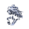
| Title | Crystal Structure of TssL from Vibrio cholerae. | ||||||
 Components Components | (VCA0115) x 2 | ||||||
 Keywords Keywords | UNKNOWN FUNCTION / T6SS / VIBRIO CHOLERAE / TSSL / VCA0115 | ||||||
| Function / homology | Type IV / VI secretion system, DotU / Type IV / VI secretion system, DotU / Type IV / VI secretion system, DotU superfamily / Type VI secretion system protein DotU / Serine Threonine Protein Phosphatase 5, Tetratricopeptide repeat / Alpha Horseshoe / Mainly Alpha / membrane / Type IV / VI secretion system DotU domain-containing protein Function and homology information Function and homology information | ||||||
| Biological species |   VIBRIO CHOLERAE (bacteria) VIBRIO CHOLERAE (bacteria) | ||||||
| Method |  X-RAY DIFFRACTION / SYNCHROTRON / MOLECULAR REPLACEMENT / Resolution: 1.499 Å X-RAY DIFFRACTION / SYNCHROTRON / MOLECULAR REPLACEMENT / Resolution: 1.499 Å | ||||||
 Authors Authors | Jeong, J.H. / Kim, Y.G. | ||||||
 Citation Citation | Journal: J. Microbiol. / Year: 2015 Title: Crystal structure of the bacterial type VI secretion system component TssL from Vibrio cholerae. Authors: Chang, J.H. / Kim, Y.G. | ||||||
| History |
|
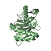
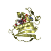
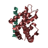
- Structure visualization
| Structure viewer | Molecule: MolmilJmol/JSmol |
|---|
- Downloads & links
-Download
| PDBx/mmCIF format | 4v3i.cif.gz | 80.7 KB | Display | PDBx/mmCIF format |
|---|---|---|---|---|
| PDB format | pdb4v3i.ent.gz | 60.7 KB | Display | PDB format |
| PDBx/mmJSON format | 4v3i.json.gz | Tree view | PDBx/mmJSON format | |
| Others |  Other downloads Other downloads |
-Validation report
| Arichive directory | https://data.pdbj.org/pub/pdb/validation_reports/v3/4v3iftp://data.pdbj.org/pub/pdb/validation_reports/v3/4v3i | HTTPS FTP |
|---|
-Related structure data
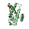
| Related structure data | 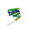 3u66S S: Starting model for refinement |
|---|---|
| Similar structure data |


-Links
 PDBj
PDBj- Assembly
| Deposited unit | 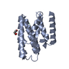
| ||||||||
|---|---|---|---|---|---|---|---|---|---|
| 1 |
| ||||||||
| Unit cell |
|
-Components
| #1: Protein | Mass: 29711.797 Da / Num. of mol.: 1 Source method: isolated from a genetically manipulated source Source: (gene. exp.) VIBRIO CHOLERAE (bacteria) / Plasmid: PET30A / Production host: |
|---|---|
| #2: Protein/peptide | Mass: 601.673 Da / Num. of mol.: 1 Source method: isolated from a genetically manipulated source Source: (gene. exp.) VIBRIO CHOLERAE (bacteria) / Plasmid: PET30A / Production host: |
| #3: Chemical | ChemComp-GOL /   Mass: 92.094 Da / Num. of mol.: 1 / Source method: obtained synthetically / Formula: C3H8O3 Mass: 92.094 Da / Num. of mol.: 1 / Source method: obtained synthetically / Formula: C3H8O3 |
| #4: Water | ChemComp-HOH /  Mass: 18.015 Da / Num. of mol.: 67 / Source method: isolated from a natural source / Formula: H2O Mass: 18.015 Da / Num. of mol.: 67 / Source method: isolated from a natural source / Formula: H2O |
-Experimental details
-Experiment
| Experiment | Method: X-RAY DIFFRACTION / Number of used crystals: 1 |
|---|
- Sample preparation
| Crystal | Density Matthews: 2.58 Å3/Da / Density % sol: 52.3 % / Description: NONE |
|---|---|
| Crystal grow | pH: 6.4 Details: 1.8 M SODIUM CHLORIDE, 0.1 M SODIUM POTASSIUM PHOSPHATE PH 6.4 |
-Data collection
| Diffraction | Mean temperature: 295 K |
|---|---|
| Diffraction source | Source: SYNCHROTRON / Site: PAL/PLS  / Beamline: 5C (4A) / Wavelength: 0.9795 / Beamline: 5C (4A) / Wavelength: 0.9795 |
| Detector | Type: ADSC QUANTUM 315 / Detector: CCD / Date: Jun 4, 2013 / Details: MIRRORS |
| Radiation | Monochromator: DOUBLE CRYSTAL MONOCHROMATER / Protocol: SINGLE WAVELENGTH / Monochromatic (M) / Laue (L): M / Scattering type: x-ray |
| Radiation wavelength | Wavelength: 0.9795 Å / Relative weight: 1 |
| Reflection | Resolution: 1.5→50 Å / Num. obs: 27803 / % possible obs: 99.8 % / Observed criterion σ(I): 0 / Redundancy: 11 % / Biso Wilson estimate: 14.82 Å2 / Rmerge(I) obs: 0.07 / Net I/σ(I): 59 |
| Reflection shell | Resolution: 1.5→1.53 Å / Redundancy: 8.5 % / Rmerge(I) obs: 0.34 / Mean I/σ(I) obs: 6.63 / % possible all: 99.9 |
- Processing
| Software |
| |||||||||||||||||||||||||||||||||||||||||||||||||||||||||||||||||||||||||||||||||||||||||||||||||||||||||
|---|---|---|---|---|---|---|---|---|---|---|---|---|---|---|---|---|---|---|---|---|---|---|---|---|---|---|---|---|---|---|---|---|---|---|---|---|---|---|---|---|---|---|---|---|---|---|---|---|---|---|---|---|---|---|---|---|---|---|---|---|---|---|---|---|---|---|---|---|---|---|---|---|---|---|---|---|---|---|---|---|---|---|---|---|---|---|---|---|---|---|---|---|---|---|---|---|---|---|---|---|---|---|---|---|---|---|
| Refinement | Method to determine structure: MOLECULAR REPLACEMENT Starting model: PDB ENTRY 3U66 Resolution: 1.499→27.979 Å / SU ML: 0.12 / σ(F): 1.58 / Phase error: 19.3 / Stereochemistry target values: ML
| |||||||||||||||||||||||||||||||||||||||||||||||||||||||||||||||||||||||||||||||||||||||||||||||||||||||||
| Solvent computation | Shrinkage radii: 0.9 Å / VDW probe radii: 1.11 Å / Solvent model: FLAT BULK SOLVENT MODEL | |||||||||||||||||||||||||||||||||||||||||||||||||||||||||||||||||||||||||||||||||||||||||||||||||||||||||
| Refinement step | Cycle: LAST / Resolution: 1.499→27.979 Å
| |||||||||||||||||||||||||||||||||||||||||||||||||||||||||||||||||||||||||||||||||||||||||||||||||||||||||
| Refine LS restraints |
| |||||||||||||||||||||||||||||||||||||||||||||||||||||||||||||||||||||||||||||||||||||||||||||||||||||||||
| LS refinement shell |
| |||||||||||||||||||||||||||||||||||||||||||||||||||||||||||||||||||||||||||||||||||||||||||||||||||||||||
| Refinement TLS params. | Method: refined / Origin x: 4.8855 Å / Origin y: 27.9566 Å / Origin z: -2.366 Å
| |||||||||||||||||||||||||||||||||||||||||||||||||||||||||||||||||||||||||||||||||||||||||||||||||||||||||
| Refinement TLS group | Selection details: ALL |
