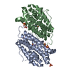
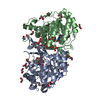
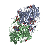
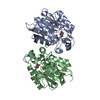
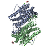
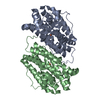
Mass: 34956.965 Da / Num. of mol.: 2 Source method: isolated from a genetically manipulated source Details: THALASSOSPIRA SP. GB04J01 THE STRAIN THALASSOSPIRA SP. GB04J01 WAS ISOLATED FROM A SEA FAN (PARAMURICEA PLACOMUS) DURING A RESEARCH-CRUISE WITH R/V HELMER HANSSEN TO THE VESTFJORDEN AREA ...Details: THALASSOSPIRA SP. GB04J01 THE STRAIN THALASSOSPIRA SP. GB04J01 WAS ISOLATED FROM A SEA FAN (PARAMURICEA PLACOMUS) DURING A RESEARCH-CRUISE WITH R/V HELMER HANSSEN TO THE VESTFJORDEN AREA (NORTHERN NORWAY AND IT IS PRESERVED AND AVAILABLE FROM AN IN-HOUSE COLLECTION AT THE UNIVERSITY OF TROMSO, NORWAY (DATABASE INDEX GB04J01). Source: (gene. exp.) THALASSOSPIRA SP. GB04J01 (bacteria) / Plasmid: PET-26B-THAEST2349 / Production host: ESCHERICHIA COLI BL21(DE3) (bacteria) / References: UniProt: A0A023T3X2, carboxylesterase
Mass: 18.015 Da / Num. of mol.: 926 / Source method: isolated from a natural source / Formula: H2O
Sequence details
THE GENE CONTAINS A N-TERMINAL LEADER SEQUENCE (27 AMINO ACIDS LONG) THAT WAS NOT INCLUDED IN THE ...THE GENE CONTAINS A N-TERMINAL LEADER SEQUENCE (27 AMINO ACIDS LONG) THAT WAS NOT INCLUDED IN THE CLONING CONSTRUCT. GB KJ365310
-
Experimental details
-
Experiment
Experiment
Method: X-RAY DIFFRACTION / Number of used crystals: 1
-
Sample preparation
Crystal
Density Matthews: 2.06 Å3/Da / Density % sol: 40.22 % / Description: NONE
Crystal grow
pH: 8 Details: THE PROTEIN WAS AT 10 MG/ML IN 50 MM TRIS-HCL PH 8.0, 500 MM NACL AND 10% GLYCEROL. RESERVOIR: 25% PEG 3350, 0.2 M MGCL2
In the structure databanks used in Yorodumi, some data are registered as the other names, "COVID-19 virus" and "2019-nCoV". Here are the details of the virus and the list of structure data.
Jan 31, 2019. EMDB accession codes are about to change! (news from PDBe EMDB page)
EMDB accession codes are about to change! (news from PDBe EMDB page)
The allocation of 4 digits for EMDB accession codes will soon come to an end. Whilst these codes will remain in use, new EMDB accession codes will include an additional digit and will expand incrementally as the available range of codes is exhausted. The current 4-digit format prefixed with “EMD-” (i.e. EMD-XXXX) will advance to a 5-digit format (i.e. EMD-XXXXX), and so on. It is currently estimated that the 4-digit codes will be depleted around Spring 2019, at which point the 5-digit format will come into force.
The EM Navigator/Yorodumi systems omit the EMD- prefix.
Related info.:Q: What is EMD? / ID/Accession-code notation in Yorodumi/EM Navigator
Yorodumi is a browser for structure data from EMDB, PDB, SASBDB, etc.
This page is also the successor to EM Navigator detail page, and also detail information page/front-end page for Omokage search.
The word "yorodu" (or yorozu) is an old Japanese word meaning "ten thousand". "mi" (miru) is to see.
Related info.:EMDB / PDB / SASBDB / Comparison of 3 databanks / Yorodumi Search / Aug 31, 2016. New EM Navigator & Yorodumi / Yorodumi Papers / Jmol/JSmol / Function and homology information / Changes in new EM Navigator and Yorodumi
 Movie
Movie Controller

 Yorodumi
 Open data
 Basic information
 Components
 Keywords
 Function and homology information
 THALASSOSPIRA SP. GB04J01 (bacteria)
 X-RAY DIFFRACTION /
  Authors
 Citation
 Structure visualization
 Downloads & links
 Other downloads
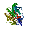


 PDBj
PDBj Assembly
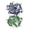

 Mass: 24.305 Da / Num. of mol.: 3 / Source method: obtained synthetically / Formula: Mg
 Mass: 18.015 Da / Num. of mol.: 926 / Source method: isolated from a natural source / Formula: H2O
 Sample preparation
 / Beamline: 14.2 / Wavelength: 0.91705
  Processing