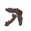
Journal: Nat Struct Mol Biol / Year: 2015 Title: A coiled-coil domain acts as a molecular ruler to regulate O-antigen chain length in lipopolysaccharide. Authors: Gregor Hagelueken / Bradley R Clarke / Hexian Huang / Anne Tuukkanen / Iulia Danciu / Dmitri I Svergun / Rohanah Hussain / Huanting Liu / Chris Whitfield / James H Naismith / Abstract: Long-chain bacterial polysaccharides have important roles in pathogenicity. In Escherichia coli O9a, a model for ABC transporter-dependent polysaccharide assembly, a large extracellular carbohydrate ...Long-chain bacterial polysaccharides have important roles in pathogenicity. In Escherichia coli O9a, a model for ABC transporter-dependent polysaccharide assembly, a large extracellular carbohydrate with a narrow size distribution is polymerized from monosaccharides by a complex of two proteins, WbdA (polymerase) and WbdD (terminating protein). Combining crystallography and small-angle X-ray scattering, we found that the C-terminal domain of WbdD contains an extended coiled-coil that physically separates WbdA from the catalytic domain of WbdD. The effects of insertions and deletions in the coiled-coil region were analyzed in vivo, revealing that polymer size is controlled by varying the length of the coiled-coil domain. Thus, the coiled-coil domain of WbdD functions as a molecular ruler that, along with WbdA:WbdD stoichiometry, controls the chain length of a model bacterial polysaccharide.
Method to determine structure: OTHER Starting model: NONE Resolution: 3.87→128.19 Å / Cor.coef. Fo:Fc: 0.909 / Cor.coef. Fo:Fc free: 0.875 / SU B: 141.891 / SU ML: 0.861 / Cross valid method: THROUGHOUT / ESU R Free: 0.916 / Stereochemistry target values: MAXIMUM LIKELIHOOD / Details: HYDROGENS HAVE BEEN ADDED IN THE RIDING POSITIONS.
 Movie
Movie Controller

 Yorodumi
 Open data
 Basic information
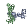
 Components
 Keywords
 Function and homology information
 X-RAY DIFFRACTION /
  Authors
 Citation


 Structure visualization
 Downloads & links
 Other downloads
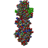

 PDBj
PDBj
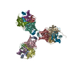
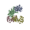
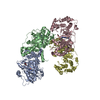
 Assembly
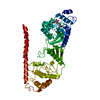

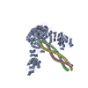
 Mass: 398.437 Da / Num. of mol.: 1 / Source method: obtained synthetically / Formula: C15H22N6O5S
 Sample preparation
 Processing