- PDB-4uut: Crystal structure of the Ultrabithorax protein -
+
Open data
ID or keywords:
Loading...
-
Basic information
Entry
Database: PDB / ID: 4uut
Title
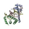
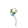
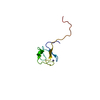
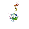
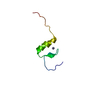
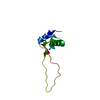
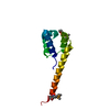
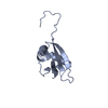
Crystal structure of the Ultrabithorax protein
Components
HOMEOTIC PROTEIN ULTRABITHORAX
Keywords
TRANSCRIPTION / UBX / HOMEOTIC PROTEIN / TRANSCRIPTION FACTOR
Function / homology
Function and homology information
dorsal vessel aortic cell fate commitment / positive regulation of muscle organ development / anterior Malpighian tubule development / specification of animal organ identity / regulation of imaginal disc growth / haltere development / mesodermal cell fate specification / specification of segmental identity, thorax / imaginal disc-derived leg morphogenesis / somatic muscle development ...dorsal vessel aortic cell fate commitment / positive regulation of muscle organ development / anterior Malpighian tubule development / specification of animal organ identity / regulation of imaginal disc growth / haltere development / mesodermal cell fate specification / specification of segmental identity, thorax / imaginal disc-derived leg morphogenesis / somatic muscle development / open tracheal system development / muscle cell fate specification / endoderm formation / polytene chromosome band / midgut development / regulation of cell fate specification / cell fate determination / anterior/posterior pattern specification / cis-regulatory region sequence-specific DNA binding / transcription repressor complex / transcription coregulator binding / protein-DNA complex / heart development / transcription regulator complex / sequence-specific DNA binding / DNA-binding transcription factor activity, RNA polymerase II-specific / RNA polymerase II cis-regulatory region sequence-specific DNA binding / protein domain specific binding / negative regulation of DNA-templated transcription / regulation of DNA-templated transcription / positive regulation of DNA-templated transcription / negative regulation of transcription by RNA polymerase II / DNA binding / nucleus Similarity search - Function
In the structure databanks used in Yorodumi, some data are registered as the other names, "COVID-19 virus" and "2019-nCoV". Here are the details of the virus and the list of structure data.
Jan 31, 2019. EMDB accession codes are about to change! (news from PDBe EMDB page)
EMDB accession codes are about to change! (news from PDBe EMDB page)
The allocation of 4 digits for EMDB accession codes will soon come to an end. Whilst these codes will remain in use, new EMDB accession codes will include an additional digit and will expand incrementally as the available range of codes is exhausted. The current 4-digit format prefixed with “EMD-” (i.e. EMD-XXXX) will advance to a 5-digit format (i.e. EMD-XXXXX), and so on. It is currently estimated that the 4-digit codes will be depleted around Spring 2019, at which point the 5-digit format will come into force.
The EM Navigator/Yorodumi systems omit the EMD- prefix.
Related info.:Q: What is EMD? / ID/Accession-code notation in Yorodumi/EM Navigator
Yorodumi is a browser for structure data from EMDB, PDB, SASBDB, etc.
This page is also the successor to EM Navigator detail page, and also detail information page/front-end page for Omokage search.
The word "yorodu" (or yorozu) is an old Japanese word meaning "ten thousand". "mi" (miru) is to see.
Related info.:EMDB / PDB / SASBDB / Comparison of 3 databanks / Yorodumi Search / Aug 31, 2016. New EM Navigator & Yorodumi / Yorodumi Papers / Jmol/JSmol / Function and homology information / Changes in new EM Navigator and Yorodumi
 Movie
Movie Controller


 Open data
 Basic information
 Components
 Keywords
 Function and homology information
 X-RAY DIFFRACTION /
  Authors
 Citation
 Structure visualization
 Downloads & links
 Other downloads

 PDBj
PDBj
 Assembly


 Mass: 96.063 Da / Num. of mol.: 4 / Source method: obtained synthetically / Formula: SO4
 Mass: 35.453 Da / Num. of mol.: 2 / Source method: obtained synthetically / Formula: Cl
 Mass: 18.015 Da / Num. of mol.: 7 / Source method: isolated from a natural source / Formula: H2O
 Sample preparation
 / Beamline: ID29 / Wavelength: 0.974716
  Processing