 Movie
Movie Controller


+ Open data

- Basic information
| Entry | Database: PDB / ID: 4uun | |||||||||
|---|---|---|---|---|---|---|---|---|---|---|
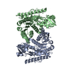
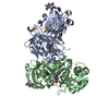
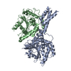
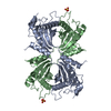
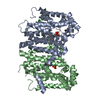
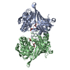
| Title | Trichomonas vaginalis lactate dehydrogenase in complex with NADH | |||||||||
 Components Components | L-LACTATE DEHYDROGENASE | |||||||||
 Keywords Keywords | OXIDOREDUCTASE | |||||||||
| Function / homology |  Function and homology information Function and homology information(S)-malate dehydrogenase (NAD+, oxaloacetate-forming) / L-malate dehydrogenase (NAD+) activity / malate metabolic process / nucleotide binding Similarity search - Function | |||||||||
| Biological species |  TRICHOMONAS VAGINALIS (eukaryote) TRICHOMONAS VAGINALIS (eukaryote) | |||||||||
| Method |  X-RAY DIFFRACTION / MOLECULAR REPLACEMENT / Resolution: 1.78 Å X-RAY DIFFRACTION / MOLECULAR REPLACEMENT / Resolution: 1.78 Å | |||||||||
 Authors Authors | Steindel, P.A. / Chen, E.H. / Theobald, D.L. | |||||||||
 Citation Citation | Journal: Protein Sci. / Year: 2016 Title: Gradual Neofunctionalization in the Convergent Evolution of Trichomonad Lactate and Malate Dehydrogenases. Authors: Steindel, P.A. / Chen, E.H. / Wirth, J.D. / Theobald, D.L. | |||||||||
| History |
|
- Structure visualization
| Structure viewer | Molecule: MolmilJmol/JSmol |
|---|
- Downloads & links
-Download
| PDBx/mmCIF format | 4uun.cif.gz | 414.6 KB | Display | PDBx/mmCIF format |
|---|---|---|---|---|
| PDB format | pdb4uun.ent.gz | 348.2 KB | Display | PDB format |
| PDBx/mmJSON format | 4uun.json.gz | Tree view | PDBx/mmJSON format | |
| Others |  Other downloads Other downloads |
-Validation report
| Arichive directory | https://data.pdbj.org/pub/pdb/validation_reports/uu/4uunftp://data.pdbj.org/pub/pdb/validation_reports/uu/4uun | HTTPS FTP |
|---|
-Related structure data
| Related structure data |  4uulC 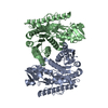 4uumC  4uuoC  4uupC 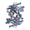 5a1tC  5mdhS C: citing same article ( S: Starting model for refinement |
|---|---|
| Similar structure data |


-Links
 PDBj
PDBj


- Assembly
| Deposited unit | 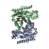
| ||||||||
|---|---|---|---|---|---|---|---|---|---|
| 1 |
| ||||||||
| Unit cell |
|
-Components
| #1: Protein | Mass: 37954.312 Da / Num. of mol.: 2 Source method: isolated from a genetically manipulated source Source: (gene. exp.) TRICHOMONAS VAGINALIS (eukaryote) / Plasmid: PET-21B / Production host:  #2: Chemical |   Mass: 665.441 Da / Num. of mol.: 2 / Source method: obtained synthetically / Formula: C21H29N7O14P2 Mass: 665.441 Da / Num. of mol.: 2 / Source method: obtained synthetically / Formula: C21H29N7O14P2#3: Water | ChemComp-HOH / |  Mass: 18.015 Da / Num. of mol.: 964 / Source method: isolated from a natural source / Formula: H2O Mass: 18.015 Da / Num. of mol.: 964 / Source method: isolated from a natural source / Formula: H2O |
|---|
-Experimental details
-Experiment
| Experiment | Method: X-RAY DIFFRACTION / Number of used crystals: 1 |
|---|
- Sample preparation
| Crystal | Density Matthews: 2.49 Å3/Da / Density % sol: 51 % / Description: NONE |
|---|---|
| Crystal grow | pH: 6.8 Details: 20 MG/ML PROTEIN, 100 MM HEPES PH 6.8, 8% PEG-6000, 5% MPD |
-Data collection
| Diffraction | Mean temperature: 100 K |
|---|---|
| Diffraction source | Source: ROTATING ANODE / Wavelength: 1.54 |
| Detector | Type: OXFORD / Detector: CCD / Date: Sep 22, 2011 |
| Radiation | Protocol: SINGLE WAVELENGTH / Monochromatic (M) / Laue (L): M / Scattering type: x-ray |
| Radiation wavelength | Wavelength: 1.54 Å / Relative weight: 1 |
| Reflection | Resolution: 1.78→46.3 Å / Num. obs: 67400 / % possible obs: 89.9 % / Observed criterion σ(I): 1.45 / Redundancy: 3.23 % / Biso Wilson estimate: 16.1 Å2 / Rmerge(I) obs: 0.071 / Net I/σ(I): 13.18 |
| Reflection shell | Resolution: 1.78→1.83 Å / Rmerge(I) obs: 0.751 / Mean I/σ(I) obs: 1.17 / % possible all: 37.5 |
- Processing
| Software |
| |||||||||||||||||||||||||||||||||||||||||||||||||||||||||||||||||||||||||||||||||||||||||||||||||||||||||||||||||||||||||||||
|---|---|---|---|---|---|---|---|---|---|---|---|---|---|---|---|---|---|---|---|---|---|---|---|---|---|---|---|---|---|---|---|---|---|---|---|---|---|---|---|---|---|---|---|---|---|---|---|---|---|---|---|---|---|---|---|---|---|---|---|---|---|---|---|---|---|---|---|---|---|---|---|---|---|---|---|---|---|---|---|---|---|---|---|---|---|---|---|---|---|---|---|---|---|---|---|---|---|---|---|---|---|---|---|---|---|---|---|---|---|---|---|---|---|---|---|---|---|---|---|---|---|---|---|---|---|---|
| Refinement | Method to determine structure: MOLECULAR REPLACEMENT Starting model: HOMOLOGY MODEL BASED ON PDB ENTRY 5MDH Resolution: 1.78→45.829 Å / SU ML: 0.18 / σ(F): 1.36 / Phase error: 20.67 / Stereochemistry target values: ML
| |||||||||||||||||||||||||||||||||||||||||||||||||||||||||||||||||||||||||||||||||||||||||||||||||||||||||||||||||||||||||||||
| Solvent computation | Shrinkage radii: 0.9 Å / VDW probe radii: 1.11 Å / Solvent model: FLAT BULK SOLVENT MODEL | |||||||||||||||||||||||||||||||||||||||||||||||||||||||||||||||||||||||||||||||||||||||||||||||||||||||||||||||||||||||||||||
| Displacement parameters | Biso mean: 27.7 Å2 | |||||||||||||||||||||||||||||||||||||||||||||||||||||||||||||||||||||||||||||||||||||||||||||||||||||||||||||||||||||||||||||
| Refinement step | Cycle: LAST / Resolution: 1.78→45.829 Å
| |||||||||||||||||||||||||||||||||||||||||||||||||||||||||||||||||||||||||||||||||||||||||||||||||||||||||||||||||||||||||||||
| Refine LS restraints |
| |||||||||||||||||||||||||||||||||||||||||||||||||||||||||||||||||||||||||||||||||||||||||||||||||||||||||||||||||||||||||||||
| LS refinement shell |
| |||||||||||||||||||||||||||||||||||||||||||||||||||||||||||||||||||||||||||||||||||||||||||||||||||||||||||||||||||||||||||||
| Refinement TLS params. | Method: refined / Refine-ID: X-RAY DIFFRACTION
| |||||||||||||||||||||||||||||||||||||||||||||||||||||||||||||||||||||||||||||||||||||||||||||||||||||||||||||||||||||||||||||
| Refinement TLS group |
|
