Protocol: SINGLE WAVELENGTH / Monochromatic (M) / Laue (L): M / Scattering type: x-ray
Radiation wavelength
Wavelength: 0.97625 Å / Relative weight: 1
Reflection
Resolution: 1.94→69.48 Å / Num. obs: 73109 / % possible obs: 99.6 % / Redundancy: 6.5 % / Rmerge(I) obs: 0.12 / Net I/σ(I): 7.5
Reflection shell
Resolution: 1.94→1.99 Å / Redundancy: 6.4 % / Rmerge(I) obs: 0.62 / Mean I/σ(I) obs: 2.2 / % possible all: 99.1
-
Processing
Software
Name
Version
Classification
xia2
datareduction
XDS
datareduction
Aimless
datascaling
SHELX
phasing
ARP/wARP
phasing
REFMAC
5.8.0073
refinement
Refinement
Method to determine structure: SAD Starting model: NONE Resolution: 1.94→69.48 Å / Cor.coef. Fo:Fc: 0.964 / Cor.coef. Fo:Fc free: 0.948 / SU B: 3.657 / SU ML: 0.103 / Cross valid method: THROUGHOUT / ESU R: 0.149 / ESU R Free: 0.144 / Stereochemistry target values: MAXIMUM LIKELIHOOD / Details: HYDROGENS HAVE BEEN ADDED IN THE RIDING POSITIONS.
Rfactor
Num. reflection
% reflection
Selection details
Rfree
0.23722
3714
5.1 %
RANDOM
Rwork
0.19334
-
-
-
obs
0.19552
69344
99.54 %
-
Solvent computation
Ion probe radii: 0.8 Å / Shrinkage radii: 0.8 Å / VDW probe radii: 1.2 Å / Solvent model: MASK
 Movie
Movie Controller


 Open data
 Basic information
 Components
 Keywords
 Function and homology information
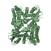
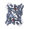
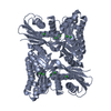
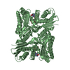
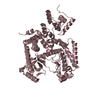
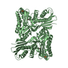
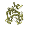
 BURKHOLDERIA PSEUDOMALLEI K96243 (bacteria)
 X-RAY DIFFRACTION /
  Authors
 Citation
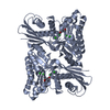
 Structure visualization
 Downloads & links
 Other downloads
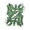


 PDBj
PDBj


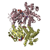
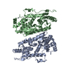
 Assembly
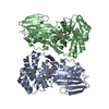

 Type: D-saccharide, alpha linking / Mass: 180.156 Da / Num. of mol.: 2
 Mass: 35.453 Da / Num. of mol.: 8 / Source method: obtained synthetically / Formula: Cl
 Mass: 18.015 Da / Num. of mol.: 797 / Source method: isolated from a natural source / Formula: H2O
 Sample preparation
 / Beamline: I03 / Wavelength: 0.97625
  Processing