Protocol: SINGLE WAVELENGTH / Monochromatic (M) / Laue (L): M / Scattering type: x-ray
Radiation wavelength
Wavelength: 1.3 Å / Relative weight: 1
Reflection twin
Crystal-ID
ID
Operator
Domain-ID
Fraction
1
1
H, K, L
1
0.722
1
1
K, H, -L
2
0.278
Reflection
Resolution: 2.46→10.99 Å / Num. obs: 55667 / % possible obs: 99.8 % / Redundancy: 5.77 % / Net I/σ(I): 25.14
-
Processing
Software
Name
Version
Classification
REFMAC
5.8.0073
refinement
PHENIX
refinement
PHENIX
modelbuilding
Coot
modelbuilding
PHENIX
phasing
Refinement
Method to determine structure: SAD / Resolution: 2.46→10.99 Å / Cor.coef. Fo:Fc: 0.957 / Cor.coef. Fo:Fc free: 0.946 / SU B: 8.868 / SU ML: 0.205 / Cross valid method: THROUGHOUT / ESU R: 0.092 / ESU R Free: 0.044 / Stereochemistry target values: MAXIMUM LIKELIHOOD / Details: HYDROGENS HAVE BEEN ADDED IN THE RIDING POSITIONS
Rfactor
Num. reflection
% reflection
Selection details
Rfree
0.18335
1461
5.3 %
RANDOM
Rwork
0.1673
-
-
-
obs
0.16814
26299
99.84 %
-
Solvent computation
Ion probe radii: 0.8 Å / Shrinkage radii: 0.8 Å / VDW probe radii: 1.2 Å / Solvent model: MASK
 Movie
Movie Controller


 Open data
 Basic information
 Components
 Keywords
 Function and homology information
 X-RAY DIFFRACTION /
  Authors
 Japan, 1items
  Citation
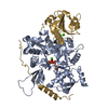
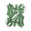
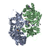
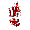
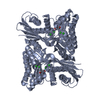
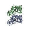
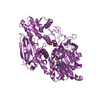
 Structure visualization
 Downloads & links
 Other downloads
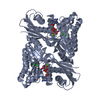

 PDBj
PDBj Assembly
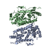


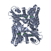
 Mass: 65.409 Da / Num. of mol.: 1 / Source method: obtained synthetically / Formula: Zn
 Mass: 18.015 Da / Num. of mol.: 67 / Source method: isolated from a natural source / Formula: H2O
 Sample preparation
 Processing