 Movie
Movie Controller

[English] 日本語
 Yorodumi
- PDB-4usv: Crystal structure of human soluble Adenylyl Cyclase with pyrophos... -
+ Open data

- Basic information
| Entry | Database: PDB / ID: 4usv | ||||||
|---|---|---|---|---|---|---|---|
| Title | Crystal structure of human soluble Adenylyl Cyclase with pyrophosphate resulting from soaking with ATP and Calcium | ||||||
 Components Components | ADENYLATE CYCLASE TYPE 10 | ||||||
 Keywords Keywords | LYASE / REACTION PRODUCT | ||||||
| Function / homology |  Function and homology information Function and homology informationnegative regulation of cardiac muscle cell contraction / mitochondrial ATP transmembrane transport / bicarbonate binding / neuron projection retraction / epithelial cilium movement involved in extracellular fluid movement / astrocyte end-foot / central region of growth cone / glucose catabolic process / positive regulation of glycogen catabolic process / regulation of mitophagy ...negative regulation of cardiac muscle cell contraction / mitochondrial ATP transmembrane transport / bicarbonate binding / neuron projection retraction / epithelial cilium movement involved in extracellular fluid movement / astrocyte end-foot / central region of growth cone / glucose catabolic process / positive regulation of glycogen catabolic process / regulation of mitophagy / regulation of membrane repolarization / adenylate cyclase / positive regulation of oxidative stress-induced neuron intrinsic apoptotic signaling pathway / basal part of cell / adenylate cyclase activity / positive regulation of ossification / cAMP biosynthetic process / neuron projection extension / : / positive regulation of vascular associated smooth muscle cell apoptotic process / positive regulation of cardiac muscle hypertrophy / positive regulation of reactive oxygen species biosynthetic process / positive regulation of mitochondrial depolarization / negative regulation of mitochondrial membrane potential / positive regulation of ATP biosynthetic process / spermatid development / positive regulation of axon extension / Hedgehog 'off' state / negative regulation of reactive oxygen species biosynthetic process / neuron projection maintenance / positive regulation of cardiac muscle cell apoptotic process / apical part of cell / adenylate cyclase-inhibiting G protein-coupled receptor signaling pathway / manganese ion binding / ATPase binding / cytoskeleton / intracellular signal transduction / cilium / neuronal cell body / dendrite / perinuclear region of cytoplasm / magnesium ion binding / mitochondrion / extracellular region / ATP binding / nucleus / plasma membrane / cytoplasm / cytosol Similarity search - Function | ||||||
| Biological species |  HOMO SAPIENS (human) HOMO SAPIENS (human) | ||||||
| Method |  X-RAY DIFFRACTION / SYNCHROTRON / MOLECULAR REPLACEMENT / Resolution: 2 Å X-RAY DIFFRACTION / SYNCHROTRON / MOLECULAR REPLACEMENT / Resolution: 2 Å | ||||||
 Authors Authors | Kleinboelting, S. / Steegborn, C. | ||||||
 Citation Citation | Journal: FEBS J. / Year: 2014 Title: Structural Analysis of Human Soluble Adenylyl Cyclase and Crystal Structures of its Nucleotide Complexes -Implications for Cyclase Catalysis and Evolution. Authors: Kleinbolting, S. / Van Den Heuvel, J. / Steegborn, C. | ||||||
| History |
|
- Structure visualization
| Structure viewer | Molecule: MolmilJmol/JSmol |
|---|
- Downloads & links
-Download
| PDBx/mmCIF format | 4usv.cif.gz | 198.9 KB | Display | PDBx/mmCIF format |
|---|---|---|---|---|
| PDB format | pdb4usv.ent.gz | 158.7 KB | Display | PDB format |
| PDBx/mmJSON format | 4usv.json.gz | Tree view | PDBx/mmJSON format | |
| Others |  Other downloads Other downloads |
-Validation report
| Arichive directory | https://data.pdbj.org/pub/pdb/validation_reports/us/4usvftp://data.pdbj.org/pub/pdb/validation_reports/us/4usv | HTTPS FTP |
|---|
-Related structure data
| Related structure data |  4ustC  4usuC  4uswC  4clkS C: citing same article ( S: Starting model for refinement |
|---|---|
| Similar structure data |


-Links
 PDBj
PDBj


- Assembly
| Deposited unit | 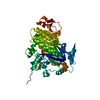
| ||||||||
|---|---|---|---|---|---|---|---|---|---|
| 1 | 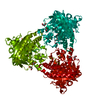
| ||||||||
| Unit cell |
|
-Components
-Protein , 1 types, 1 molecules A
| #1: Protein | Mass: 54269.664 Da / Num. of mol.: 1 / Fragment: CATALYTIC DOMAIN, RESIDUES 1-469 Source method: isolated from a genetically manipulated source Details: BETA-MERCAPTOETHANOL MODIFICATION AT RESIDUE CYS253 Source: (gene. exp.) HOMO SAPIENS (human) / Plasmid: PVL1392 / Cell line (production host): High Five / Production host:  TRICHOPLUSIA NI (cabbage looper) / References: UniProt: Q96PN6, adenylate cyclase TRICHOPLUSIA NI (cabbage looper) / References: UniProt: Q96PN6, adenylate cyclase |
|---|
-Non-polymers , 7 types, 192 molecules 


| #2: Chemical | ChemComp-CL /  Mass: 35.453 Da / Num. of mol.: 1 / Source method: obtained synthetically / Formula: Cl Mass: 35.453 Da / Num. of mol.: 1 / Source method: obtained synthetically / Formula: Cl | ||||||||
|---|---|---|---|---|---|---|---|---|---|
| #3: Chemical | ChemComp-CA /  Mass: 40.078 Da / Num. of mol.: 1 / Source method: obtained synthetically / Formula: Ca Mass: 40.078 Da / Num. of mol.: 1 / Source method: obtained synthetically / Formula: Ca | ||||||||
| #4: Chemical |  Mass: 62.068 Da / Num. of mol.: 2 / Source method: obtained synthetically / Formula: C2H6O2 Mass: 62.068 Da / Num. of mol.: 2 / Source method: obtained synthetically / Formula: C2H6O2#5: Chemical | ChemComp-ACT / |  Mass: 59.044 Da / Num. of mol.: 1 / Source method: obtained synthetically / Formula: C2H3O2 Mass: 59.044 Da / Num. of mol.: 1 / Source method: obtained synthetically / Formula: C2H3O2#6: Chemical | ChemComp-POP / |  Mass: 175.959 Da / Num. of mol.: 1 / Source method: obtained synthetically / Formula: H2O7P2 Mass: 175.959 Da / Num. of mol.: 1 / Source method: obtained synthetically / Formula: H2O7P2#7: Chemical | ChemComp-GOL / |  Mass: 92.094 Da / Num. of mol.: 1 / Source method: obtained synthetically / Formula: C3H8O3 Mass: 92.094 Da / Num. of mol.: 1 / Source method: obtained synthetically / Formula: C3H8O3#8: Water | ChemComp-HOH / | Mass: 18.015 Da / Num. of mol.: 185 / Source method: isolated from a natural source / Formula: H2O |
-Details
| Has protein modification | Y |
|---|
-Experimental details
-Experiment
| Experiment | Method: X-RAY DIFFRACTION / Number of used crystals: 1 |
|---|
- Sample preparation
| Crystal | Density Matthews: 2.4 Å3/Da / Density % sol: 54 % / Description: NONE |
|---|---|
| Crystal grow | pH: 6 Details: 0.1 M SODIUM ACETATE PH 4.8, 0.2 M TRI-SODIUM-CITRATE, 15% (W/V) PEG 4000, 10% (V/V) GLYCEROL |
-Data collection
| Diffraction | Mean temperature: 100 K |
|---|---|
| Diffraction source | Source: SYNCHROTRON / Site: BESSY  / Beamline: 14.1 / Wavelength: 0.91705 / Beamline: 14.1 / Wavelength: 0.91705 |
| Detector | Type: DECTRIS PILATUS 6M / Detector: PIXEL / Date: Jul 5, 2013 / Details: COLLIMATOR |
| Radiation | Monochromator: SI111-DCM WITH SAGITTAL BENDER / Protocol: SINGLE WAVELENGTH / Monochromatic (M) / Laue (L): M / Scattering type: x-ray |
| Radiation wavelength | Wavelength: 0.91705 Å / Relative weight: 1 |
| Reflection | Resolution: 2→87.36 Å / Num. obs: 37919 / % possible obs: 100 % / Observed criterion σ(I): 2 / Redundancy: 6.3 % / Rmerge(I) obs: 0.07 / Net I/σ(I): 18.11 |
| Reflection shell | Resolution: 2→2.05 Å / Redundancy: 6.5 % / Rmerge(I) obs: 0.8 / Mean I/σ(I) obs: 2.2 / % possible all: 100 |
- Processing
| Software |
| ||||||||||||||||||||||||||||||||||||||||||||||||||||||||||||||||||||||||||||||||||||||||||||||||||||||||||||||||||||||||||||||||||||||||||||||||||||||||||||||||||||||||||||||||||||||
|---|---|---|---|---|---|---|---|---|---|---|---|---|---|---|---|---|---|---|---|---|---|---|---|---|---|---|---|---|---|---|---|---|---|---|---|---|---|---|---|---|---|---|---|---|---|---|---|---|---|---|---|---|---|---|---|---|---|---|---|---|---|---|---|---|---|---|---|---|---|---|---|---|---|---|---|---|---|---|---|---|---|---|---|---|---|---|---|---|---|---|---|---|---|---|---|---|---|---|---|---|---|---|---|---|---|---|---|---|---|---|---|---|---|---|---|---|---|---|---|---|---|---|---|---|---|---|---|---|---|---|---|---|---|---|---|---|---|---|---|---|---|---|---|---|---|---|---|---|---|---|---|---|---|---|---|---|---|---|---|---|---|---|---|---|---|---|---|---|---|---|---|---|---|---|---|---|---|---|---|---|---|---|---|
| Refinement | Method to determine structure: MOLECULAR REPLACEMENT Starting model: PDB ENTRY 4CLK Resolution: 2→87.36 Å / Cor.coef. Fo:Fc: 0.971 / Cor.coef. Fo:Fc free: 0.947 / SU B: 7.852 / SU ML: 0.106 / Cross valid method: THROUGHOUT / ESU R: 0.141 / ESU R Free: 0.139 / Stereochemistry target values: MAXIMUM LIKELIHOOD Details: HYDROGENS HAVE BEEN ADDED IN THE RIDING POSITIONS. U VALUES WITH TLS ADDED
| ||||||||||||||||||||||||||||||||||||||||||||||||||||||||||||||||||||||||||||||||||||||||||||||||||||||||||||||||||||||||||||||||||||||||||||||||||||||||||||||||||||||||||||||||||||||
| Solvent computation | Ion probe radii: 0.8 Å / Shrinkage radii: 0.8 Å / VDW probe radii: 1.2 Å / Solvent model: MASK | ||||||||||||||||||||||||||||||||||||||||||||||||||||||||||||||||||||||||||||||||||||||||||||||||||||||||||||||||||||||||||||||||||||||||||||||||||||||||||||||||||||||||||||||||||||||
| Displacement parameters | Biso mean: 43.485 Å2
| ||||||||||||||||||||||||||||||||||||||||||||||||||||||||||||||||||||||||||||||||||||||||||||||||||||||||||||||||||||||||||||||||||||||||||||||||||||||||||||||||||||||||||||||||||||||
| Refinement step | Cycle: LAST / Resolution: 2→87.36 Å
| ||||||||||||||||||||||||||||||||||||||||||||||||||||||||||||||||||||||||||||||||||||||||||||||||||||||||||||||||||||||||||||||||||||||||||||||||||||||||||||||||||||||||||||||||||||||
| Refine LS restraints |
|