 Movie
Movie Controller

[English] 日本語
 Yorodumi
- PDB-4upu: Crystal structure of IP3 3-K calmodulin binding region in complex... -
+ Open data

- Basic information
| Entry | Database: PDB / ID: 4upu | ||||||
|---|---|---|---|---|---|---|---|
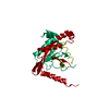
| Title | Crystal structure of IP3 3-K calmodulin binding region in complex with Calmodulin | ||||||
 Components Components |
| ||||||
 Keywords Keywords | TRANSFERASE | ||||||
| Function / homology |  Function and homology information Function and homology informationinositol-trisphosphate 3-kinase / inositol-1,4,5-trisphosphate 3-kinase activity / inositol hexakisphosphate kinase activity / inositol phosphate biosynthetic process / modification of postsynaptic actin cytoskeleton / : / : / : / : / positive regulation of protein autophosphorylation ...inositol-trisphosphate 3-kinase / inositol-1,4,5-trisphosphate 3-kinase activity / inositol hexakisphosphate kinase activity / inositol phosphate biosynthetic process / modification of postsynaptic actin cytoskeleton / : / : / : / : / positive regulation of protein autophosphorylation / : / negative regulation of peptidyl-threonine phosphorylation / : / type 3 metabotropic glutamate receptor binding / postsynaptic actin cytoskeleton / calcium/calmodulin-dependent protein kinase activity / positive regulation of DNA binding / CaM pathway / Cam-PDE 1 activation / positive regulation of peptidyl-threonine phosphorylation / Sodium/Calcium exchangers / Calmodulin induced events / Reduction of cytosolic Ca++ levels / Activation of Ca-permeable Kainate Receptor / CREB1 phosphorylation through the activation of CaMKII/CaMKK/CaMKIV cascasde / Loss of phosphorylation of MECP2 at T308 / CREB1 phosphorylation through the activation of Adenylate Cyclase / negative regulation of high voltage-gated calcium channel activity / PKA activation / CaMK IV-mediated phosphorylation of CREB / Glycogen breakdown (glycogenolysis) / response to corticosterone / negative regulation of ryanodine-sensitive calcium-release channel activity / Activation of RAC1 downstream of NMDARs / organelle localization by membrane tethering / CLEC7A (Dectin-1) induces NFAT activation / : / autophagosome membrane docking / regulation of synaptic vesicle exocytosis / negative regulation of calcium ion export across plasma membrane / regulation of cardiac muscle cell action potential / presynaptic endocytosis / phosphatidylinositol phosphate biosynthetic process / Synthesis of IP3 and IP4 in the cytosol / positive regulation of protein serine/threonine kinase activity / Phase 0 - rapid depolarisation / Negative regulation of NMDA receptor-mediated neuronal transmission / Unblocking of NMDA receptors, glutamate binding and activation / calcineurin-mediated signaling / RHO GTPases activate PAKs / nitric-oxide synthase binding / regulation of cell communication by electrical coupling involved in cardiac conduction / Ion transport by P-type ATPases / adenylate cyclase binding / Uptake and function of anthrax toxins / protein phosphatase activator activity / regulation of ryanodine-sensitive calcium-release channel activity / Long-term potentiation / Calcineurin activates NFAT / Regulation of MECP2 expression and activity / DARPP-32 events / Smooth Muscle Contraction / regulation of synaptic vesicle endocytosis / detection of calcium ion / catalytic complex / regulation of cardiac muscle contraction / cellular response to interferon-beta / RHO GTPases activate IQGAPs / positive regulation of nitric-oxide synthase activity / phosphatidylinositol 3-kinase binding / activation of adenylate cyclase activity / calcium channel inhibitor activity / presynaptic cytosol / Activation of AMPK downstream of NMDARs / regulation of release of sequestered calcium ion into cytosol by sarcoplasmic reticulum / enzyme regulator activity / eNOS activation / Ion homeostasis / Tetrahydrobiopterin (BH4) synthesis, recycling, salvage and regulation / regulation of calcium-mediated signaling / Protein methylation / titin binding / regulation of cardiac muscle contraction by regulation of the release of sequestered calcium ion / voltage-gated potassium channel complex / FCERI mediated Ca+2 mobilization / calcium channel complex / substantia nigra development / regulation of heart rate / FCGR3A-mediated IL10 synthesis / cellular response to calcium ion / calyx of Held / Ras activation upon Ca2+ influx through NMDA receptor / Antigen activates B Cell Receptor (BCR) leading to generation of second messengers / response to amphetamine / nitric-oxide synthase regulator activity / adenylate cyclase activator activity / protein serine/threonine kinase activator activity / VEGFR2 mediated cell proliferation / VEGFR2 mediated vascular permeability / regulation of cytokinesis Similarity search - Function | ||||||
| Biological species |  HOMO SAPIENS (human) HOMO SAPIENS (human) | ||||||
| Method |  X-RAY DIFFRACTION / SYNCHROTRON / MOLECULAR REPLACEMENT / Resolution: 2.34 Å X-RAY DIFFRACTION / SYNCHROTRON / MOLECULAR REPLACEMENT / Resolution: 2.34 Å | ||||||
 Authors Authors | Franco-Echevarria, E. / Banos-Sanz, J.I. / Monterroso, B. / Round, A. / Sanz-Aparicio, J. / Gonzalez, B. | ||||||
 Citation Citation | Journal: Biochem.J. / Year: 2014 Title: A New Calmodulin Binding Motif for Inositol 1,4,5-Trisphosphate 3-Kinase Regulation. Authors: Franco-Echevarria, E. / Banos-Sanz, J.I. / Monterroso, B. / Round, A. / Sanz-Aparicio, J. / Gonzalez, B. | ||||||
| History |
|
- Structure visualization
| Structure viewer | Molecule: MolmilJmol/JSmol |
|---|
- Downloads & links
-Download
| PDBx/mmCIF format | 4upu.cif.gz | 50.4 KB | Display | PDBx/mmCIF format |
|---|---|---|---|---|
| PDB format | pdb4upu.ent.gz | 36.3 KB | Display | PDB format |
| PDBx/mmJSON format | 4upu.json.gz | Tree view | PDBx/mmJSON format | |
| Others |  Other downloads Other downloads |
-Validation report
| Arichive directory | https://data.pdbj.org/pub/pdb/validation_reports/up/4upuftp://data.pdbj.org/pub/pdb/validation_reports/up/4upu | HTTPS FTP |
|---|
-Related structure data
| Related structure data |  2vayS S: Starting model for refinement |
|---|---|
| Similar structure data |


-Links
 PDBj
PDBj


- Assembly
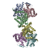
| Deposited unit | 
| ||||||||||||
|---|---|---|---|---|---|---|---|---|---|---|---|---|---|
| 1 | x 24
| ||||||||||||
| Unit cell |
| ||||||||||||
| Components on special symmetry positions |
|
-Components
| #1: Protein | Mass: 16721.350 Da / Num. of mol.: 1 / Fragment: RESIDUES 2-149 Source method: isolated from a genetically manipulated source Source: (gene. exp.) HOMO SAPIENS (human) / Production host:  | ||||
|---|---|---|---|---|---|
| #2: Protein/peptide | Mass: 3027.520 Da / Num. of mol.: 1 / Fragment: CALMODULIN BINDING REGION, RESIDUES 158-183 Source method: isolated from a genetically manipulated source Source: (gene. exp.) HOMO SAPIENS (human) / Plasmid: PKLSLT / Production host: References: UniProt: P23677, inositol-trisphosphate 3-kinase | ||||
| #3: Chemical | ChemComp-CA /   Mass: 40.078 Da / Num. of mol.: 5 / Source method: obtained synthetically / Formula: Ca Mass: 40.078 Da / Num. of mol.: 5 / Source method: obtained synthetically / Formula: Ca#4: Chemical | ChemComp-GOL / |   Mass: 92.094 Da / Num. of mol.: 1 / Source method: obtained synthetically / Formula: C3H8O3 Mass: 92.094 Da / Num. of mol.: 1 / Source method: obtained synthetically / Formula: C3H8O3#5: Water | ChemComp-HOH / |  Mass: 18.015 Da / Num. of mol.: 81 / Source method: isolated from a natural source / Formula: H2O Mass: 18.015 Da / Num. of mol.: 81 / Source method: isolated from a natural source / Formula: H2O |
-Experimental details
-Experiment
| Experiment | Method: X-RAY DIFFRACTION / Number of used crystals: 1 |
|---|
- Sample preparation
| Crystal | Density Matthews: 4 Å3/Da / Density % sol: 69 % / Description: NONE |
|---|---|
| Crystal grow | pH: 4 Details: 29% W/V POLYETHYLENE GLYCOL 4000, 0.2 M (NH4)2SO4, 0.1 M SODIUM ACETATE TRIHYDRATE PH 4.0 AND 10 MM SPERMIDINE |
-Data collection
| Diffraction | Mean temperature: 100 K |
|---|---|
| Diffraction source | Source: SYNCHROTRON / Site: ALBA  / Beamline: XALOC / Wavelength: 0.97949 / Beamline: XALOC / Wavelength: 0.97949 |
| Detector | Type: DECTRIS PILATUS 6M / Detector: PIXEL / Date: Oct 29, 2012 / Details: KB FOCUSING MIRRORS |
| Radiation | Protocol: SINGLE WAVELENGTH / Monochromatic (M) / Laue (L): M / Scattering type: x-ray |
| Radiation wavelength | Wavelength: 0.97949 Å / Relative weight: 1 |
| Reflection | Resolution: 2.34→79.74 Å / Num. obs: 14994 / % possible obs: 100 % / Observed criterion σ(I): 2 / Redundancy: 13.9 % / Rmerge(I) obs: 0.09 / Net I/σ(I): 5.91 |
| Reflection shell | Resolution: 2.34→2.46 Å / Redundancy: 12.66 % / Rmerge(I) obs: 0.35 / Mean I/σ(I) obs: 2.06 / % possible all: 100 |
- Processing
| Software |
| ||||||||||||||||||||||||||||||||||||||||||||||||||||||||||||||||||||||||||||||||||||||||||||||||||||||||||||||||||||||||||||||||||||||||||||||||||||||||||||||||||||||||||||||||||||||
|---|---|---|---|---|---|---|---|---|---|---|---|---|---|---|---|---|---|---|---|---|---|---|---|---|---|---|---|---|---|---|---|---|---|---|---|---|---|---|---|---|---|---|---|---|---|---|---|---|---|---|---|---|---|---|---|---|---|---|---|---|---|---|---|---|---|---|---|---|---|---|---|---|---|---|---|---|---|---|---|---|---|---|---|---|---|---|---|---|---|---|---|---|---|---|---|---|---|---|---|---|---|---|---|---|---|---|---|---|---|---|---|---|---|---|---|---|---|---|---|---|---|---|---|---|---|---|---|---|---|---|---|---|---|---|---|---|---|---|---|---|---|---|---|---|---|---|---|---|---|---|---|---|---|---|---|---|---|---|---|---|---|---|---|---|---|---|---|---|---|---|---|---|---|---|---|---|---|---|---|---|---|---|---|
| Refinement | Method to determine structure: MOLECULAR REPLACEMENT Starting model: PDB ENTRY 2VAY Resolution: 2.34→79.65 Å / Cor.coef. Fo:Fc: 0.945 / Cor.coef. Fo:Fc free: 0.935 / SU B: 3.581 / SU ML: 0.087 / Cross valid method: THROUGHOUT / ESU R: 0.039 / ESU R Free: 0.034 / Stereochemistry target values: MAXIMUM LIKELIHOOD Details: HYDROGENS HAVE BEEN ADDED IN THE RIDING POSITIONS. HYDROGENS HAVE BEEN USED IF PRESENT IN THE INPUT U VALUES REFINED INDIVIDUALLY
| ||||||||||||||||||||||||||||||||||||||||||||||||||||||||||||||||||||||||||||||||||||||||||||||||||||||||||||||||||||||||||||||||||||||||||||||||||||||||||||||||||||||||||||||||||||||
| Solvent computation | Ion probe radii: 0.8 Å / Shrinkage radii: 0.8 Å / VDW probe radii: 1.2 Å / Solvent model: MASK | ||||||||||||||||||||||||||||||||||||||||||||||||||||||||||||||||||||||||||||||||||||||||||||||||||||||||||||||||||||||||||||||||||||||||||||||||||||||||||||||||||||||||||||||||||||||
| Displacement parameters | Biso mean: 35.465 Å2
| ||||||||||||||||||||||||||||||||||||||||||||||||||||||||||||||||||||||||||||||||||||||||||||||||||||||||||||||||||||||||||||||||||||||||||||||||||||||||||||||||||||||||||||||||||||||
| Refinement step | Cycle: LAST / Resolution: 2.34→79.65 Å
| ||||||||||||||||||||||||||||||||||||||||||||||||||||||||||||||||||||||||||||||||||||||||||||||||||||||||||||||||||||||||||||||||||||||||||||||||||||||||||||||||||||||||||||||||||||||
| Refine LS restraints |
|