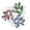
nucleoside-diphosphate kinase / UTP biosynthetic process / CTP biosynthetic process / nucleoside diphosphate kinase activity / GTP biosynthetic process / ATP binding / metal ion binding Similarity search - Function
Mass: 18.015 Da / Num. of mol.: 419 / Source method: isolated from a natural source / Formula: H2O
-
Experimental details
-
Experiment
Experiment
Method: X-RAY DIFFRACTION / Number of used crystals: 1
-
Sample preparation
Crystal
Density Matthews: 2.1 Å3/Da / Density % sol: 42.5 % Description: THE STARTING MODEL WAS PREVIOUS LITOPENAEUS VANNAMEI NDK STRUCTURE COMPLEXED WITH DADP WHICH WILL BE DEPOSITED IN PDB
Crystal grow
pH: 8.5 Details: 0.2 M MAGNESIUM CHLORIDE HEXAHYDRATE, 0.1 M TRIS-HCL PH 8.5 AND 30% (W/V) PEG 4,000
Monochromator: SI(111) CHANNEL CUT / Protocol: SINGLE WAVELENGTH / Monochromatic (M) / Laue (L): M / Scattering type: x-ray
Radiation wavelength
Wavelength: 0.979 Å / Relative weight: 1
Reflection
Resolution: 2→34.8 Å / Num. obs: 29462 / % possible obs: 99.6 % / Observed criterion σ(I): 3 / Redundancy: 8.2 % / Rmerge(I) obs: 0.14 / Net I/σ(I): 16.7
Reflection shell
Resolution: 2→2.1 Å / Redundancy: 8.1 % / Rmerge(I) obs: 0.74 / Mean I/σ(I) obs: 3.4 / % possible all: 96.4
-
Processing
Software
Name
Version
Classification
PHENIX
(PHENIX.REFINE: 1.8.2_1309)
refinement
HKL-2000
datareduction
SCALEPACK
datascaling
PHASER
phasing
Refinement
Method to determine structure: MOLECULAR REPLACEMENT Starting model: SHRIMP NDK STRUCTURE TO BE DEPOSITED Resolution: 2.007→34.887 Å / SU ML: 0.18 / σ(F): 1.34 / Phase error: 24.19 / Stereochemistry target values: ML Details: NO ELECTRON DENSITY WAS FOUND FOR ADP IN THE CHAIN A ACTIVE SITE. ALSO NUCLEOTIDE BINDING LOOP IN CHAIN A THAT COMPRISE RESIDUES 44-66 WERE DISORDERED. THEREFORE, BOTH LIGAND AND DISORDERED ...Details: NO ELECTRON DENSITY WAS FOUND FOR ADP IN THE CHAIN A ACTIVE SITE. ALSO NUCLEOTIDE BINDING LOOP IN CHAIN A THAT COMPRISE RESIDUES 44-66 WERE DISORDERED. THEREFORE, BOTH LIGAND AND DISORDERED LOOP WERE NOT INCLUDED IN IN FINAL THE MODEL.
Rfactor
Num. reflection
% reflection
Rfree
0.217
1497
5.1 %
Rwork
0.178
-
-
obs
0.18
29447
99.58 %
Solvent computation
Shrinkage radii: 0.9 Å / VDW probe radii: 1.11 Å / Solvent model: FLAT BULK SOLVENT MODEL
Refinement step
Cycle: LAST / Resolution: 2.007→34.887 Å
Protein
Nucleic acid
Ligand
Solvent
Total
Num. atoms
3371
0
56
419
3846
Refine LS restraints
Refine-ID
Type
Dev ideal
Number
X-RAY DIFFRACTION
f_bond_d
0.007
3493
X-RAY DIFFRACTION
f_angle_d
1.169
4720
X-RAY DIFFRACTION
f_dihedral_angle_d
14.657
1302
X-RAY DIFFRACTION
f_chiral_restr
0.081
503
X-RAY DIFFRACTION
f_plane_restr
0.004
594
LS refinement shell
Resolution (Å)
Rfactor Rfree
Num. reflection Rfree
Rfactor Rwork
Num. reflection Rwork
Refine-ID
% reflection obs (%)
2.0072-2.0719
0.2987
128
0.2411
2423
X-RAY DIFFRACTION
96
2.0719-2.146
0.2755
131
0.2079
2510
X-RAY DIFFRACTION
100
2.146-2.2319
0.2225
127
0.1941
2524
X-RAY DIFFRACTION
100
2.2319-2.3334
0.2522
130
0.1993
2538
X-RAY DIFFRACTION
100
2.3334-2.4564
0.2415
134
0.1906
2513
X-RAY DIFFRACTION
100
2.4564-2.6103
0.2478
124
0.1904
2543
X-RAY DIFFRACTION
100
2.6103-2.8117
0.2431
143
0.1903
2533
X-RAY DIFFRACTION
100
2.8117-3.0945
0.2335
153
0.1767
2525
X-RAY DIFFRACTION
100
3.0945-3.542
0.2201
119
0.1614
2602
X-RAY DIFFRACTION
100
3.542-4.461
0.1704
152
0.1537
2569
X-RAY DIFFRACTION
100
4.461-34.8925
0.1951
156
0.1712
2670
X-RAY DIFFRACTION
100
+
About Yorodumi
-
News
-
Feb 9, 2022. New format data for meta-information of EMDB entries
New format data for meta-information of EMDB entries
Version 3 of the EMDB header file is now the official format.
The previous official version 1.9 will be removed from the archive.
In the structure databanks used in Yorodumi, some data are registered as the other names, "COVID-19 virus" and "2019-nCoV". Here are the details of the virus and the list of structure data.
Jan 31, 2019. EMDB accession codes are about to change! (news from PDBe EMDB page)
EMDB accession codes are about to change! (news from PDBe EMDB page)
The allocation of 4 digits for EMDB accession codes will soon come to an end. Whilst these codes will remain in use, new EMDB accession codes will include an additional digit and will expand incrementally as the available range of codes is exhausted. The current 4-digit format prefixed with “EMD-” (i.e. EMD-XXXX) will advance to a 5-digit format (i.e. EMD-XXXXX), and so on. It is currently estimated that the 4-digit codes will be depleted around Spring 2019, at which point the 5-digit format will come into force.
The EM Navigator/Yorodumi systems omit the EMD- prefix.
Related info.:Q: What is EMD? / ID/Accession-code notation in Yorodumi/EM Navigator
Yorodumi is a browser for structure data from EMDB, PDB, SASBDB, etc.
This page is also the successor to EM Navigator detail page, and also detail information page/front-end page for Omokage search.
The word "yorodu" (or yorozu) is an old Japanese word meaning "ten thousand". "mi" (miru) is to see.
Related info.:EMDB / PDB / SASBDB / Comparison of 3 databanks / Yorodumi Search / Aug 31, 2016. New EM Navigator & Yorodumi / Yorodumi Papers / Jmol/JSmol / Function and homology information / Changes in new EM Navigator and Yorodumi
 Movie
Movie Controller

 Yorodumi
 Open data
 Basic information
 Components
 Keywords
 Function and homology information
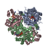
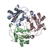
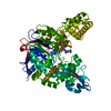
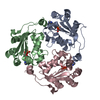
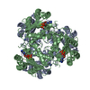
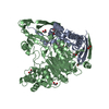
 LITOPENAEUS VANNAMEI (Pacific white shrimp)
 X-RAY DIFFRACTION /
  Authors
 Citation
 Structure visualization
 Downloads & links
 Other downloads
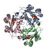
 PDBj
PDBj Assembly


 Mass: 427.201 Da / Num. of mol.: 2 / Source method: obtained synthetically / Formula: C10H15N5O10P2 / Comment: ADP, energy-carrying molecule*YM
 Mass: 24.305 Da / Num. of mol.: 2 / Source method: obtained synthetically / Formula: Mg
 Mass: 18.015 Da / Num. of mol.: 419 / Source method: isolated from a natural source / Formula: H2O
 Sample preparation
 / Beamline: X6A / Wavelength: 0.979
  Processing