 Movie
Movie Controller

[English] 日本語
 Yorodumi
- PDB-4uog: Crystallographic structure of nucleoside diphosphate kinase from ... -
+ Open data

- Basic information
| Entry | Database: PDB / ID: 4uog | ||||||
|---|---|---|---|---|---|---|---|
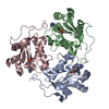
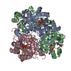
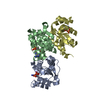
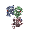
| Title | Crystallographic structure of nucleoside diphosphate kinase from Litopenaeus vannamei complexed with dCDP | ||||||
 Components Components | NUCLEOSIDE DIPHOSPHATE KINASE | ||||||
 Keywords Keywords | TRANSFERASE / BINARY / COMPLEX / DCDP / PYRIMIDINE / BINDING SITE / DESOXYNUCLEOTIDE | ||||||
| Function / homology |  Function and homology information Function and homology informationnucleoside-diphosphate kinase / UTP biosynthetic process / CTP biosynthetic process / nucleoside diphosphate kinase activity / GTP biosynthetic process / ATP binding / metal ion binding Similarity search - Function | ||||||
| Biological species |  LITOPENAEUS VANNAMEI (Pacific white shrimp) LITOPENAEUS VANNAMEI (Pacific white shrimp) | ||||||
| Method |  X-RAY DIFFRACTION / SYNCHROTRON / MOLECULAR REPLACEMENT / Resolution: 2.3 Å X-RAY DIFFRACTION / SYNCHROTRON / MOLECULAR REPLACEMENT / Resolution: 2.3 Å | ||||||
 Authors Authors | Lopez-Zavala, A.A. / Stojanoff, V. / Rudino-Pinera, E. / Sotelo-Mundo, R.R. | ||||||
 Citation Citation | Journal: Acta Crystallogr.,Sect.F / Year: 2014 Title: Structure of Nucleoside Diphosphate Kinase from Pacific Shrimp (Litopenaeus Vannamei) in Binary Complexes with Purine and Pyrimidine Nucleoside Diphosphates Authors: Lopez-Zavala, A.A. / Quintero-Reyes, I.E. / Carrasco-Miranda, J.S. / Stojanoff, V. / Weichsel, A. / Rudino-Pinera, E. / Sotelo-Mundo, R.R. | ||||||
| History |
|
- Structure visualization
| Structure viewer | Molecule: MolmilJmol/JSmol |
|---|
- Downloads & links
-Download
| PDBx/mmCIF format | 4uog.cif.gz | 109.1 KB | Display | PDBx/mmCIF format |
|---|---|---|---|---|
| PDB format | pdb4uog.ent.gz | 85.2 KB | Display | PDB format |
| PDBx/mmJSON format | 4uog.json.gz | Tree view | PDBx/mmJSON format | |
| Others |  Other downloads Other downloads |
-Validation report
| Arichive directory | https://data.pdbj.org/pub/pdb/validation_reports/uo/4uogftp://data.pdbj.org/pub/pdb/validation_reports/uo/4uog | HTTPS FTP |
|---|
-Related structure data
| Related structure data | 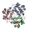 4uofSC 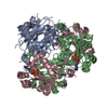 4uohC S: Starting model for refinement C: citing same article ( |
|---|---|
| Similar structure data |

-Links
 PDBj
PDBj- Assembly
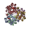
| Deposited unit | 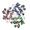
| ||||||||
|---|---|---|---|---|---|---|---|---|---|
| 1 | 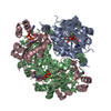
| ||||||||
| Unit cell |
|
-Components
| #1: Protein | Mass: 17015.627 Da / Num. of mol.: 3 Source method: isolated from a genetically manipulated source Source: (gene. exp.) LITOPENAEUS VANNAMEI (Pacific white shrimp)Plasmid: PJEXPRESS414 / Production host:  #2: Chemical |   Mass: 387.177 Da / Num. of mol.: 3 / Source method: obtained synthetically / Formula: C9H15N3O10P2 Mass: 387.177 Da / Num. of mol.: 3 / Source method: obtained synthetically / Formula: C9H15N3O10P2#3: Chemical |   Mass: 24.305 Da / Num. of mol.: 3 / Source method: obtained synthetically / Formula: Mg Mass: 24.305 Da / Num. of mol.: 3 / Source method: obtained synthetically / Formula: Mg#4: Water | ChemComp-HOH / |  Mass: 18.015 Da / Num. of mol.: 319 / Source method: isolated from a natural source / Formula: H2O Mass: 18.015 Da / Num. of mol.: 319 / Source method: isolated from a natural source / Formula: H2O |
|---|
-Experimental details
-Experiment
| Experiment | Method: X-RAY DIFFRACTION / Number of used crystals: 1 |
|---|
- Sample preparation
| Crystal | Density Matthews: 2.4 Å3/Da / Density % sol: 49.6 % / Description: NONE |
|---|---|
| Crystal grow | pH: 8.5 Details: 0.2 M AMMONIUM ACETATE, 0.1 M TRIS-HCL PH 8.5 AND 30% (V/V) 2-PROPANOL |
-Data collection
| Diffraction | Mean temperature: 100 K |
|---|---|
| Diffraction source | Source: SYNCHROTRON / Site: NSLS  / Beamline: X6A / Wavelength: 0.979 / Beamline: X6A / Wavelength: 0.979 |
| Detector | Type: ADSC QUANTUM 270 / Detector: CCD / Date: Nov 1, 2013 / Details: TOROIDAL FOCUSING MIRROR |
| Radiation | Monochromator: SI(111) CHANNEL CUT / Protocol: SINGLE WAVELENGTH / Monochromatic (M) / Laue (L): M / Scattering type: x-ray |
| Radiation wavelength | Wavelength: 0.979 Å / Relative weight: 1 |
| Reflection | Resolution: 2.3→19.3 Å / Num. obs: 21500 / % possible obs: 95 % / Observed criterion σ(I): 3 / Redundancy: 8.2 % / Biso Wilson estimate: 22.5 Å2 / Rmerge(I) obs: 0.17 / Net I/σ(I): 13.4 |
| Reflection shell | Resolution: 2.3→2.4 Å / Redundancy: 8.3 % / Rmerge(I) obs: 0.7 / Mean I/σ(I) obs: 3.5 / % possible all: 97.7 |
- Processing
| Software |
| |||||||||||||||||||||||||||||||||||||||||||||||||||||||||||||||
|---|---|---|---|---|---|---|---|---|---|---|---|---|---|---|---|---|---|---|---|---|---|---|---|---|---|---|---|---|---|---|---|---|---|---|---|---|---|---|---|---|---|---|---|---|---|---|---|---|---|---|---|---|---|---|---|---|---|---|---|---|---|---|---|---|
| Refinement | Method to determine structure: MOLECULAR REPLACEMENT Starting model: PDB ENTRY 4UOF Resolution: 2.3→19.304 Å / SU ML: 0.33 / σ(F): 1.34 / Phase error: 24.96 / Stereochemistry target values: ML
| |||||||||||||||||||||||||||||||||||||||||||||||||||||||||||||||
| Solvent computation | Shrinkage radii: 0.9 Å / VDW probe radii: 1.11 Å / Solvent model: FLAT BULK SOLVENT MODEL / Bsol: 25 Å2 | |||||||||||||||||||||||||||||||||||||||||||||||||||||||||||||||
| Displacement parameters | Biso mean: 21.8 Å2 | |||||||||||||||||||||||||||||||||||||||||||||||||||||||||||||||
| Refinement step | Cycle: LAST / Resolution: 2.3→19.304 Å
| |||||||||||||||||||||||||||||||||||||||||||||||||||||||||||||||
| Refine LS restraints |
| |||||||||||||||||||||||||||||||||||||||||||||||||||||||||||||||
| LS refinement shell |
|
