 Movie
Movie Controller


+ Open data

- Basic information
| Entry | Database: PDB / ID: 4uis | ||||||
|---|---|---|---|---|---|---|---|
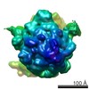
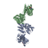
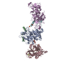
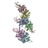
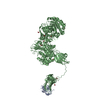
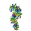
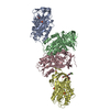
| Title | The cryoEM structure of human gamma-Secretase complex | ||||||
 Components Components |
| ||||||
 Keywords Keywords | HYDROLASE / GAMMA-SECRETASE | ||||||
| Function / homology |  Function and homology information Function and homology informationCajal-Retzius cell differentiation / positive regulation of L-glutamate import across plasma membrane / amyloid precursor protein biosynthetic process / gamma-secretase complex / aspartic endopeptidase activity, intramembrane cleaving / positive regulation of amyloid precursor protein biosynthetic process / smooth endoplasmic reticulum calcium ion homeostasis / Noncanonical activation of NOTCH3 / protein catabolic process at postsynapse / TGFBR3 PTM regulation ...Cajal-Retzius cell differentiation / positive regulation of L-glutamate import across plasma membrane / amyloid precursor protein biosynthetic process / gamma-secretase complex / aspartic endopeptidase activity, intramembrane cleaving / positive regulation of amyloid precursor protein biosynthetic process / smooth endoplasmic reticulum calcium ion homeostasis / Noncanonical activation of NOTCH3 / protein catabolic process at postsynapse / TGFBR3 PTM regulation / : / : / Notch receptor processing / positive regulation of coagulation / negative regulation of axonogenesis / membrane protein intracellular domain proteolysis / short-term synaptic potentiation / skin morphogenesis / T cell activation involved in immune response / choline transport / central nervous system myelination / NOTCH4 Activation and Transmission of Signal to the Nucleus / ciliary rootlet / regulation of resting membrane potential / neural retina development / Regulated proteolysis of p75NTR / myeloid dendritic cell differentiation / L-glutamate import across plasma membrane / endoplasmic reticulum calcium ion homeostasis / brain morphogenesis / amyloid precursor protein metabolic process / regulation of long-term synaptic potentiation / locomotion / cell fate specification / astrocyte activation involved in immune response / regulation of synaptic vesicle cycle / embryonic limb morphogenesis / regulation of postsynapse organization / myeloid cell homeostasis / regulation of canonical Wnt signaling pathway / aggresome / G protein-coupled dopamine receptor signaling pathway / skeletal system morphogenesis / Hydrolases; Acting on peptide bonds (peptidases); Aspartic endopeptidases / growth factor receptor binding / azurophil granule membrane / positive regulation of amyloid fibril formation / dorsal/ventral neural tube patterning / glutamate receptor signaling pathway / amyloid-beta formation / amyloid precursor protein catabolic process / mitochondrial transport / blood vessel development / heart looping / regulation of neuron projection development / positive regulation of dendritic spine development / cerebral cortex cell migration / smooth endoplasmic reticulum / membrane protein ectodomain proteolysis / adult behavior / positive regulation of receptor recycling / nuclear outer membrane / negative regulation of epidermal growth factor receptor signaling pathway / negative regulation of apoptotic signaling pathway / EPH-ephrin mediated repulsion of cells / T cell proliferation / hematopoietic progenitor cell differentiation / Nuclear signaling by ERBB4 / somitogenesis / viral release from host cell by cytolysis / negative regulation of ubiquitin-dependent protein catabolic process / neuron development / endopeptidase activator activity / autophagosome assembly / regulation of synaptic transmission, glutamatergic / calcium ion homeostasis / Degradation of the extracellular matrix / Notch signaling pathway / peptidoglycan catabolic process / neuron projection maintenance / rough endoplasmic reticulum / epithelial cell proliferation / astrocyte activation / cellular response to calcium ion / NOTCH2 Activation and Transmission of Signal to the Nucleus / cerebellum development / thymus development / positive regulation of glycolytic process / NRIF signals cell death from the nucleus / Activated NOTCH1 Transmits Signal to the Nucleus / dendritic shaft / post-embryonic development / PDZ domain binding / neuromuscular junction / NOTCH3 Activation and Transmission of Signal to the Nucleus / apoptotic signaling pathway / neuron cellular homeostasis / protein processing / cell-cell adhesion / sarcolemma Similarity search - Function | ||||||
| Biological species |  HOMO SAPIENS (human) HOMO SAPIENS (human) | ||||||
| Method | ELECTRON MICROSCOPY / single particle reconstruction / cryo EM / Resolution: 4.4 Å | ||||||
 Authors Authors | Sun, L. / Zhao, L. / Yang, G. / Yan, C. / Zhou, R. / Zhou, X. / Xie, T. / Zhao, Y. / Wu, S. / Li, X. / Shi, Y. | ||||||
 Citation Citation | Journal: Proc Natl Acad Sci U S A / Year: 2015 Title: Structural basis of human γ-secretase assembly. Authors: Linfeng Sun / Lingyun Zhao / Guanghui Yang / Chuangye Yan / Rui Zhou / Xiaoyuan Zhou / Tian Xie / Yanyu Zhao / Shenjie Wu / Xueming Li / Yigong Shi /  Abstract: The four-component intramembrane protease γ-secretase is intricately linked to the development of Alzheimer's disease. Despite recent structural advances, the transmembrane segments (TMs) of γ- ...The four-component intramembrane protease γ-secretase is intricately linked to the development of Alzheimer's disease. Despite recent structural advances, the transmembrane segments (TMs) of γ-secretase remain to be specifically assigned. Here we report a 3D structure of human γ-secretase at 4.32-Å resolution, determined by single-particle, electron cryomicroscopy in the presence of digitonin and with a T4 lysozyme fused to the amino terminus of presenilin 1 (PS1). The overall structure of this human γ-secretase is very similar to that of wild-type γ-secretase determined in the presence of amphipols. The 20 TMs are unambiguously assigned to the four components, revealing principles of subunit assembly. Within the transmembrane region, PS1 is centrally located, with its amino-terminal fragment (NTF) packing against Pen-2 and its carboxyl-terminal fragment (CTF) interacting with Aph-1. The only TM of nicastrin associates with Aph-1 at the thick end of the TM horseshoe, and the extracellular domain of nicastrin directly binds Pen-2 at the thin end. TM6 and TM7 in PS1, which harbor the catalytic aspartate residues, are located on the convex side of the TM horseshoe. This structure serves as an important framework for understanding the function and mechanism of γ-secretase. | ||||||
| History |
|
- Structure visualization
| Movie |
Movie viewer |
|---|---|
| Structure viewer | Molecule: MolmilJmol/JSmol |
- Downloads & links
-Download
| PDBx/mmCIF format | 4uis.cif.gz | 286.4 KB | Display | PDBx/mmCIF format |
|---|---|---|---|---|
| PDB format | pdb4uis.ent.gz | 230.5 KB | Display | PDB format |
| PDBx/mmJSON format | 4uis.json.gz | Tree view | PDBx/mmJSON format | |
| Others |  Other downloads Other downloads |
-Validation report
| Arichive directory | https://data.pdbj.org/pub/pdb/validation_reports/ui/4uisftp://data.pdbj.org/pub/pdb/validation_reports/ui/4uis | HTTPS FTP |
|---|
-Related structure data


-Links
 PDBj
PDBj


- Assembly
| Deposited unit | 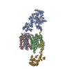
|
|---|---|
| 1 |
|
-Components
| #1: Protein | Mass: 63331.977 Da / Num. of mol.: 1 Source method: isolated from a genetically manipulated source Source: (gene. exp.) HOMO SAPIENS (human) / Cell line (production host): HEK 293S / Production host: HOMO SAPIENS (human) / References: UniProt: Q92542*PLUS |
|---|---|
| #2: Protein | Mass: 23547.639 Da / Num. of mol.: 1 Source method: isolated from a genetically manipulated source Source: (gene. exp.) HOMO SAPIENS (human) / Cell line (production host): HEK 293S / Production host: HOMO SAPIENS (human) / References: UniProt: P49768*PLUS |
| #3: Protein | Mass: 16698.539 Da / Num. of mol.: 1 Source method: isolated from a genetically manipulated source Source: (gene. exp.) HOMO SAPIENS (human) / Cell line (production host): HEK 293S / Production host: HOMO SAPIENS (human) |
| #4: Protein | Mass: 5294.518 Da / Num. of mol.: 1 Source method: isolated from a genetically manipulated source Source: (gene. exp.) HOMO SAPIENS (human) / Cell line (production host): HEK 293S / Production host: HOMO SAPIENS (human) |
| #5: Protein | Mass: 18435.207 Da / Num. of mol.: 1 Source method: isolated from a genetically manipulated source Source: (gene. exp.) HOMO SAPIENS (human) / Cell line (production host): HEK 293S / Production host: HOMO SAPIENS (human)References: UniProt: A0A097J809, UniProt: D9IEF7*PLUS, lysozyme |
| Has protein modification | Y |
-Experimental details
-Experiment
| Experiment | Method: ELECTRON MICROSCOPY |
|---|---|
| EM experiment | Aggregation state: PARTICLE / 3D reconstruction method: single particle reconstruction |
- Sample preparation
| Component | Name: T4-LYSOZYME FUSION GAMMA- SECRETASE / Type: COMPLEX |
|---|---|
| Buffer solution | Name: 0.1% DIGITONIN, 25 MM HEPES, PH 7.4, AND 150 MM NACL. / pH: 7.4 Details: 0.1% DIGITONIN, 25 MM HEPES, PH 7.4, AND 150 MM NACL. |
| Specimen | Conc.: 4.2 mg/ml / Embedding applied: NO / Shadowing applied: NO / Staining applied: NO / Vitrification applied: YES |
| Specimen support | Details: HOLEY CARBON |
| Vitrification | Instrument: FEI VITROBOT MARK IV / Cryogen name: ETHANE Details: VITRIFICATION 1 -- CRYOGEN- ETHANE, HUMIDITY- 100, TEMPERATURE- 277, INSTRUMENT- FEI VITROBOT MARK IV, METHOD- BLOT FOR 3 SECONDS BEFORE PLUNGING, |
- Electron microscopy imaging
| Experimental equipment |  Model: Titan Krios / Image courtesy: FEI Company |
|---|---|
| Microscopy | Model: FEI TITAN KRIOS / Date: Dec 22, 2014 |
| Electron gun | Electron source:  FIELD EMISSION GUN / Accelerating voltage: 300 kV / Illumination mode: SPOT SCAN FIELD EMISSION GUN / Accelerating voltage: 300 kV / Illumination mode: SPOT SCAN |
| Electron lens | Mode: BRIGHT FIELD / Nominal defocus max: 3000 nm / Nominal defocus min: 1500 nm / Cs: 1.4 mm |
| Image recording | Electron dose: 4.5 e/Å2 / Film or detector model: DIRECT ELECTRON DE-12 (4k x 3k) |
| Image scans | Num. digital images: 2000 |
- Processing
| EM software |
| ||||||||||||
|---|---|---|---|---|---|---|---|---|---|---|---|---|---|
| Symmetry | Point symmetry: C1 (asymmetric) | ||||||||||||
| 3D reconstruction | Resolution: 4.4 Å / Num. of particles: 177207 / Nominal pixel size: 1.32 Å / Actual pixel size: 1.32 Å Details: SUBMISSION BASED ON EXPERIMENTAL DATA FROM EMDB EMD-2974. (DEPOSITION ID: 13293 Symmetry type: POINT | ||||||||||||
| Atomic model building | Protocol: FLEXIBLE FIT / Space: REAL / Details: METHOD--FLEXIBLE | ||||||||||||
| Atomic model building | PDB-ID: 4R12 Accession code: 4R12 / Source name: PDB / Type: experimental model | ||||||||||||
| Refinement | Highest resolution: 4.4 Å | ||||||||||||
| Refinement step | Cycle: LAST / Highest resolution: 4.4 Å
|

