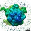
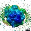
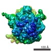
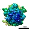
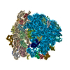
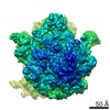
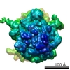
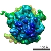
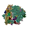
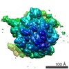
Journal: Structure / Year: 2015 Title: Structural dynamics of ribosome subunit association studied by mixing-spraying time-resolved cryogenic electron microscopy. Authors: Bo Chen / Sandip Kaledhonkar / Ming Sun / Bingxin Shen / Zonghuan Lu / David Barnard / Toh-Ming Lu / Ruben L Gonzalez / Joachim Frank / Abstract: Ribosomal subunit association is a key checkpoint in translation initiation but its structural dynamics are poorly understood. Here, we used a recently developed mixing-spraying, time-resolved, ...Ribosomal subunit association is a key checkpoint in translation initiation but its structural dynamics are poorly understood. Here, we used a recently developed mixing-spraying, time-resolved, cryogenic electron microscopy (cryo-EM) method to study ribosomal subunit association in the sub-second time range. We have improved this method and increased the cryo-EM data yield by tenfold. Pre-equilibrium states of the association reaction were captured by reacting the mixture of ribosomal subunits for 60 ms and 140 ms. We also identified three distinct ribosome conformations in the associated ribosomes. The observed proportions of these conformations are the same in these two time points, suggesting that ribosomes equilibrate among the three conformations within less than 60 ms upon formation. Our results demonstrate that the mixing-spraying method can capture multiple states of macromolecules during a sub-second reaction. Other fast processes, such as translation initiation, decoding, and ribosome recycling, are amenable to study with this method.
History
Deposition
Apr 7, 2015
-
Header (metadata) release
Jun 17, 2015
-
Map release
Jun 17, 2015
-
Update
Jul 1, 2015
-
Current status
Jul 1, 2015
Processing site: PDBe / Status: Released
-
Structure visualization
Movie
Surface view with section colored by density value
Cryogen name: ETHANE / Chamber humidity: 80 % / Chamber temperature: 80 K / Instrument: OTHER Details: Equal volume of 1.2 microM 30S and 0.6 microM 50S (final concentration after mixing) were injected into the mixing-spraying device each at flow rate of 3 microL/s. The computer-controlled ...Details: Equal volume of 1.2 microM 30S and 0.6 microM 50S (final concentration after mixing) were injected into the mixing-spraying device each at flow rate of 3 microL/s. The computer-controlled plunging device was purchased from Dr. Howard White (Eastern Virginia Medical School, VA). Timed resolved state: Vitrified after spraying
-
Electron microscopy
Microscope
FEI TECNAI F20
Temperature
Average: 80 K
Details
Low dose, Data was collected over two years time
Date
Sep 13, 2013
Image recording
Category: CCD / Film or detector model: GATAN ULTRASCAN 4000 (4k x 4k) / Number real images: 3402 / Average electron dose: 17 e/Å2
Electron beam
Acceleration voltage: 200 kV / Electron source: FIELD EMISSION GUN
In the structure databanks used in Yorodumi, some data are registered as the other names, "COVID-19 virus" and "2019-nCoV". Here are the details of the virus and the list of structure data.
Jan 31, 2019. EMDB accession codes are about to change! (news from PDBe EMDB page)
EMDB accession codes are about to change! (news from PDBe EMDB page)
The allocation of 4 digits for EMDB accession codes will soon come to an end. Whilst these codes will remain in use, new EMDB accession codes will include an additional digit and will expand incrementally as the available range of codes is exhausted. The current 4-digit format prefixed with “EMD-” (i.e. EMD-XXXX) will advance to a 5-digit format (i.e. EMD-XXXXX), and so on. It is currently estimated that the 4-digit codes will be depleted around Spring 2019, at which point the 5-digit format will come into force.
The EM Navigator/Yorodumi systems omit the EMD- prefix.
Related info.:Q: What is EMD? / ID/Accession-code notation in Yorodumi/EM Navigator
Yorodumi is a browser for structure data from EMDB, PDB, SASBDB, etc.
This page is also the successor to EM Navigator detail page, and also detail information page/front-end page for Omokage search.
The word "yorodu" (or yorozu) is an old Japanese word meaning "ten thousand". "mi" (miru) is to see.
Related info.:EMDB / PDB / SASBDB / Comparison of 3 databanks / Yorodumi Search / Aug 31, 2016. New EM Navigator & Yorodumi / Yorodumi Papers / Jmol/JSmol / Function and homology information / Changes in new EM Navigator and Yorodumi
 Movie
Movie Controller

 Yorodumi
 Open data
 Basic information
 Map data
 Sample
 Keywords
 Function and homology information
 Authors
 Citation
 Structure visualization
 Downloads & links
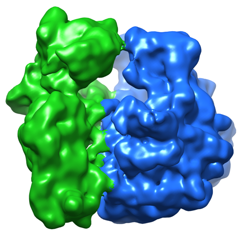 emd_2978.png
 http://ftp.pdbj.org/pub/emdb/structures/EMD-2978


 Z (Sec.)
 Y (Row.)
 X (Col.)


 Sample components
 Processing
 Electron microscopy
 FIELD EMISSION GUN
