[English] 日本語
Yorodumi
- PDB-4ozq: Crystal structure of the mouse Kif14 motor domain -

+
Open data


ID or keywords:

Loading...

-
Basic information

Entry
Database: PDB / ID: 4ozq
TitleCrystal structure of the mouse Kif14 motor domain
ComponentsChimera of Maltose-binding periplasmic protein and Kinesin family member 14 protein
KeywordsMOTOR PROTEIN / Kinesin / ATPase
Function / homology
Function and homology information


cerebellar granular layer structural organization / regulation of cell maturation / cerebellar Purkinje cell layer structural organization / RHO GTPases activate CIT / RND2 GTPase cycle / negative regulation of integrin activation / RND1 GTPase cycle / cell proliferation in forebrain / regulation of Rap protein signal transduction / cerebellar cortex development ...cerebellar granular layer structural organization / regulation of cell maturation / cerebellar Purkinje cell layer structural organization / RHO GTPases activate CIT / RND2 GTPase cycle / negative regulation of integrin activation / RND1 GTPase cycle / cell proliferation in forebrain / regulation of Rap protein signal transduction / cerebellar cortex development / olfactory bulb development / plus-end-directed microtubule motor activity / Flemming body / SCF-dependent proteasomal ubiquitin-dependent protein catabolic process / regulation of myelination / kinesin complex / microtubule motor activity / mitotic metaphase chromosome alignment / microtubule-based movement / positive regulation of cytokinesis / activation of protein kinase activity / regulation of G1/S transition of mitotic cell cycle / spindle midzone / regulation of cell adhesion / regulation of neuron apoptotic process / regulation of cell migration / regulation of G2/M transition of mitotic cell cycle / tubulin binding / substrate adhesion-dependent cell spreading / PDZ domain binding / regulation of cell growth / hippocampus development / establishment of protein localization / cerebral cortex development / midbody / microtubule binding / proteasome-mediated ubiquitin-dependent protein catabolic process / negative regulation of neuron apoptotic process / microtubule / cell division / positive regulation of cell population proliferation / negative regulation of apoptotic process / protein kinase binding / ATP hydrolysis activity / ATP binding / membrane / nucleus / plasma membrane / cytosol
Similarity search - Function
Kinesin-associated / Kinesin-associated / Forkhead associated domain / Forkhead-associated (FHA) domain profile. / FHA domain / Forkhead-associated (FHA) domain / Kinesin motor domain signature. / Kinesin motor domain, conserved site / Kinesin motor domain / Kinesin motor domain profile. ...Kinesin-associated / Kinesin-associated / Forkhead associated domain / Forkhead-associated (FHA) domain profile. / FHA domain / Forkhead-associated (FHA) domain / Kinesin motor domain signature. / Kinesin motor domain, conserved site / Kinesin motor domain / Kinesin motor domain profile. / Kinesin motor, catalytic domain. ATPase. / Kinesin motor domain / SMAD/FHA domain superfamily / Kinesin motor domain superfamily / P-loop containing nucleoside triphosphate hydrolase
Similarity search - Domain/homology
ADENOSINE-5'-DIPHOSPHATE / Kinesin-like protein KIF14 / :
Similarity search - Component
Biological speciesEscherichia coli UMEA 3304-1 (bacteria)
Mus musculus (house mouse)
MethodX-RAY DIFFRACTION / SYNCHROTRON / Resolution: 2.71 Å
AuthorsArora, K. / Talje, L. / Asenjo, A.B. / Andersen, P. / Atchia, K. / Joshi, M. / Sosa, H. / Kwok, B.H. / Allingham, J.S.
Funding support Canada, 1items
OrganizationGrant numberCountry
Canadian Institutes of Health Canada
Citation
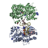
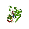
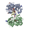
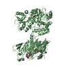
Journal: J Mol Biol / Year: 2014
Title: KIF14 binds tightly to microtubules and adopts a rigor-like conformation.
Authors: Kritica Arora / Lama Talje / Ana B Asenjo / Parker Andersen / Kaleem Atchia / Monika Joshi / Hernando Sosa / John S Allingham / Benjamin H Kwok /
Abstract: The mitotic kinesin motor protein KIF14 is essential for cytokinesis during cell division and has been implicated in cerebral development and a variety of human cancers. Here we show that the mouse ...The mitotic kinesin motor protein KIF14 is essential for cytokinesis during cell division and has been implicated in cerebral development and a variety of human cancers. Here we show that the mouse KIF14 motor domain binds tightly to microtubules and does not display typical nucleotide-dependent changes in this affinity. It also has robust ATPase activity but very slow motility. A crystal structure of the ADP-bound form of the KIF14 motor domain reveals a dramatically opened ATP-binding pocket, as if ready to exchange its bound ADP for Mg·ATP. In this state, the central β-sheet is twisted ~10° beyond the maximal amount observed in other kinesins. This configuration has only been seen in the nucleotide-free states of myosins-known as the "rigor-like" state. Fitting of this atomic model to electron density maps from cryo-electron microscopy indicates a distinct binding configuration of the motor domain to microtubules. We postulate that these properties of KIF14 are well suited for stabilizing midbody microtubules during cytokinesis.
History
DepositionFeb 18, 2014Deposition site: RCSB / Processing site: RCSB
Revision 1.0Jul 9, 2014Provider: repository / Type: Initial release
Revision 1.1Oct 1, 2014Group: Database references
Revision 1.2Feb 4, 2015Group: Derived calculations
Revision 1.3Dec 27, 2023Group: Advisory / Data collection ...Advisory / Data collection / Database references / Derived calculations / Other / Refinement description / Source and taxonomy
Category: chem_comp_atom / chem_comp_bond ...chem_comp_atom / chem_comp_bond / citation / database_2 / entity_src_gen / pdbx_database_status / pdbx_prerelease_seq / pdbx_struct_assembly / pdbx_struct_oper_list / pdbx_validate_polymer_linkage / refine_hist / struct_ncs_dom_lim / struct_ref
Item: _citation.journal_id_CSD / _database_2.pdbx_DOI ..._citation.journal_id_CSD / _database_2.pdbx_DOI / _database_2.pdbx_database_accession / _entity_src_gen.pdbx_alt_source_flag / _pdbx_database_status.pdb_format_compatible / _pdbx_struct_assembly.oligomeric_details / _pdbx_struct_oper_list.symmetry_operation / _refine_hist.pdbx_number_atoms_nucleic_acid / _refine_hist.pdbx_number_atoms_protein / _struct_ncs_dom_lim.beg_auth_comp_id / _struct_ncs_dom_lim.beg_label_asym_id / _struct_ncs_dom_lim.beg_label_comp_id / _struct_ncs_dom_lim.beg_label_seq_id / _struct_ncs_dom_lim.end_auth_comp_id / _struct_ncs_dom_lim.end_label_asym_id / _struct_ncs_dom_lim.end_label_comp_id / _struct_ncs_dom_lim.end_label_seq_id / _struct_ref.biol_id

-
Structure visualization

Structure viewerMolecule:
MolmilJmol/JSmol

Downloads & links

-
Assembly

Deposited unit
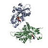
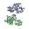
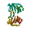
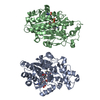
A: Chimera of Maltose-binding periplasmic protein and Kinesin family member 14 protein
B: Chimera of Maltose-binding periplasmic protein and Kinesin family member 14 protein
hetero molecules


Theoretical massNumber of molelcules
Total (without water)159,9306
Polymers158,9512
Non-polymers9794
Water64936
1
A: Chimera of Maltose-binding periplasmic protein and Kinesin family member 14 protein
hetero molecules


Theoretical massNumber of molelcules
Total (without water)79,9653
Polymers79,4761
Non-polymers4892
Water181
TypeNameSymmetry operationNumber
identity operation1_555x,y,z1
MethodPISA
2
B: Chimera of Maltose-binding periplasmic protein and Kinesin family member 14 protein
hetero molecules


Theoretical massNumber of molelcules
Total (without water)79,9653
Polymers79,4761
Non-polymers4892
Water181
TypeNameSymmetry operationNumber
identity operation1_555x,y,z1
MethodPISA
Unit cell
Length a, b, c (Å)65.137, 72.193, 94.098
Angle α, β, γ (deg.)74.430, 87.450, 89.940
Int Tables number1
Space group name H-MP1
Noncrystallographic symmetry (NCS)NCS domain:
IDEns-IDDetails
11A
21B

NCS domain segments:

Component-ID: 0 / Ens-ID: 1 / Beg auth comp-ID: TRP / Beg label comp-ID: TRP / End auth comp-ID: ARG / End label comp-ID: ARG / Refine code: 0 / Auth seq-ID: -360 - 734 / Label seq-ID: 11 - 716

Dom-IDAuth asym-IDLabel asym-ID
1AA
2BB

-
Components

#1: Protein Chimera of Maltose-binding periplasmic protein and Kinesin family member 14 protein


Mass: 79475.672 Da / Num. of mol.: 2
Source method: isolated from a genetically manipulated source
Source: (gene. exp.) Escherichia coli UMEA 3304-1 (bacteria), (gene. exp.) Mus musculus (house mouse)
Gene: G962_03763, Kif14,kif14 / Production host: Escherichia coli (E. coli) / References: UniProt: T9HUW4, UniProt: L0N7N1
#2: Chemical ChemComp-ADP / ADENOSINE-5'-DIPHOSPHATE / Adenosine diphosphate


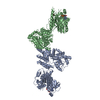
Mass: 427.201 Da / Num. of mol.: 2 / Source method: obtained synthetically / Formula: C10H15N5O10P2 / Comment: ADP, energy-carrying molecule*YM
#3: Chemical ChemComp-EDO / 1,2-ETHANEDIOL / ETHYLENE GLYCOL / Ethylene glycol


Mass: 62.068 Da / Num. of mol.: 2 / Source method: obtained synthetically / Formula: C2H6O2
#4: Water ChemComp-HOH / water / Water


Mass: 18.015 Da / Num. of mol.: 36 / Source method: isolated from a natural source / Formula: H2O

-
Experimental details

-
Experiment

ExperimentMethod: X-RAY DIFFRACTION

-
Sample preparation

CrystalDensity Matthews: 2.81 Å3/Da / Density % sol: 56.28 %
Crystal growTemperature: 277 K / Method: vapor diffusion, hanging drop / pH: 7 / Details: PEG 3350, sodium formate

-
Data collection

DiffractionMean temperature: 93 K
Diffraction sourceSource: SYNCHROTRON / Site: NSLS / Beamline: X6A / Wavelength: 1.0781 Å
DetectorType: ADSC QUANTUM 270 / Detector: CCD / Date: Oct 25, 2013
RadiationProtocol: SINGLE WAVELENGTH / Scattering type: x-ray
Radiation wavelengthWavelength: 1.0781 Å / Relative weight: 1
ReflectionResolution: 2.71→30 Å / Num. obs: 40225 / % possible obs: 94.6 % / Redundancy: 3.1 % / Net I/σ(I): 10.72

-
Processing

SoftwareName: REFMAC / Version: 5.7.0032 / Classification: refinement
RefinementResolution: 2.71→29.36 Å / Cor.coef. Fo:Fc: 0.883 / Cor.coef. Fo:Fc free: 0.829 / SU B: 39.339 / SU ML: 0.387 / Cross valid method: THROUGHOUT / σ(F): 0 / ESU R Free: 0.456 / Stereochemistry target values: MAXIMUM LIKELIHOOD
Details: HYDROGENS HAVE BEEN ADDED IN THE RIDING POSITIONS U VALUES : WITH TLS ADDED
RfactorNum. reflection% reflectionSelection details
Rfree0.3075 2034 5.1 %RANDOM
Rwork0.2638 38158 --
obs0.2661 40192 89.5 %-
Solvent computationIon probe radii: 0.8 Å / Shrinkage radii: 0.8 Å / VDW probe radii: 1.2 Å / Solvent model: MASK
Displacement parametersBiso max: 122.54 Å2 / Biso mean: 46.597 Å2 / Biso min: 13.18 Å2
Baniso -1Baniso -2Baniso -3
1--3.07 Å2-0.55 Å2-0.45 Å2
2--1.3 Å2-0.38 Å2
3---1.41 Å2
Refinement stepCycle: final / Resolution: 2.71→29.36 Å
ProteinNucleic acidLigandSolventTotal
Num. atoms10166 0 62 36 10264
Biso mean--18.16 18.75 -
Num. residues----1370
Refine LS restraints
Refine-IDTypeDev idealDev ideal targetNumber
X-RAY DIFFRACTIONr_bond_refined_d0.0110.01910454
X-RAY DIFFRACTIONr_bond_other_d0.0030.029490
X-RAY DIFFRACTIONr_angle_refined_deg1.4721.95714264
X-RAY DIFFRACTIONr_angle_other_deg1.379321759
X-RAY DIFFRACTIONr_dihedral_angle_1_deg6.37451358
X-RAY DIFFRACTIONr_dihedral_angle_2_deg36.88124.915413
X-RAY DIFFRACTIONr_dihedral_angle_3_deg17.909151527
X-RAY DIFFRACTIONr_dihedral_angle_4_deg18.6311530
X-RAY DIFFRACTIONr_chiral_restr0.0790.21630
X-RAY DIFFRACTIONr_gen_planes_refined0.0060.02112033
X-RAY DIFFRACTIONr_gen_planes_other0.0030.022327
X-RAY DIFFRACTIONr_mcbond_it0.2490.3695468
X-RAY DIFFRACTIONr_mcbond_other0.2480.3695467
X-RAY DIFFRACTIONr_mcangle_it0.3960.5536814
Refine LS restraints NCS

Ens-ID: 1 / Number: 35484 / Refine-ID: X-RAY DIFFRACTION / Type: interatomic distance / Rms dev position: 0.11 Å / Weight position: 0.05

Dom-IDAuth asym-ID
1A
2B
LS refinement shellResolution: 2.708→2.779 Å / Total num. of bins used: 20
RfactorNum. reflection% reflection
Rfree0.447 59 -
Rwork0.385 977 -
all-1036 -
obs--30.91 %
Refinement TLS params.

Method: refined / Refine-ID: X-RAY DIFFRACTION

IDL112)L122)L132)L222)L232)L332)S11 (Å °)S12 (Å °)S13 (Å °)S21 (Å °)S22 (Å °)S23 (Å °)S31 (Å °)S32 (Å °)S33 (Å °)T112)T122)T132)T222)T232)T332)Origin x (Å)Origin y (Å)Origin z (Å)
11.3411-0.33911.17711.8711-0.58812.8459-0.1627-0.14140.15680.3854-0.0134-0.3497-0.46060.17840.17610.6064-0.0705-0.19250.17750.17420.66068.27911.991-10.386
22.15220.3894-0.42432.5869-0.71191.16020.00820.0819-0.2285-0.05780.02680.0930.1055-0.0528-0.03490.4303-0.0306-0.10390.01010.04120.590721.9339.103-53.564
31.7372-0.22110.12181.32680.10111.928-0.08540.3691-0.0272-0.22910.0193-0.07880.26080.36680.06620.5718-0.0924-0.10080.25620.16620.5915-18.573-12.56711.963
41.8481-0.5590.84932.7129-0.491.3371-0.1029-0.17720.14080.21680.0051-0.0048-0.1862-0.02850.09780.4316-0.0523-0.08250.03530.06480.6172-6.366-9.25954.894
Refinement TLS group
IDRefine-IDRefine TLS-IDAuth asym-IDAuth seq-ID
1X-RAY DIFFRACTION1A-361 - 0
2X-RAY DIFFRACTION2A390 - 734
3X-RAY DIFFRACTION3B-368 - 0
4X-RAY DIFFRACTION4B390 - 734

+
About Yorodumi

-
News

-
Feb 9, 2022. New format data for meta-information of EMDB entries

New format data for meta-information of EMDB entries

  • Version 3 of the EMDB header file is now the official format.
  • The previous official version 1.9 will be removed from the archive.

Related info.:EMDB header

External links:wwPDB to switch to version 3 of the EMDB data model

-
Aug 12, 2020. Covid-19 info

Covid-19 info

URL: https://pdbj.org/emnavi/covid19.php

New page: Covid-19 featured information page in EM Navigator.

Related info.:Covid-19 info / Mar 5, 2020. Novel coronavirus structure data

+
Mar 5, 2020. Novel coronavirus structure data

Novel coronavirus structure data

Related info.:Yorodumi Speices / Aug 12, 2020. Covid-19 info

External links:COVID-19 featured content - PDBj / Molecule of the Month (242):Coronavirus Proteases

+
Jan 31, 2019. EMDB accession codes are about to change! (news from PDBe EMDB page)

EMDB accession codes are about to change! (news from PDBe EMDB page)

  • The allocation of 4 digits for EMDB accession codes will soon come to an end. Whilst these codes will remain in use, new EMDB accession codes will include an additional digit and will expand incrementally as the available range of codes is exhausted. The current 4-digit format prefixed with “EMD-” (i.e. EMD-XXXX) will advance to a 5-digit format (i.e. EMD-XXXXX), and so on. It is currently estimated that the 4-digit codes will be depleted around Spring 2019, at which point the 5-digit format will come into force.
  • The EM Navigator/Yorodumi systems omit the EMD- prefix.

Related info.:Q: What is EMD? / ID/Accession-code notation in Yorodumi/EM Navigator

External links:EMDB Accession Codes are Changing Soon! / Contact to PDBj

+
Jul 12, 2017. Major update of PDB

Major update of PDB

  • wwPDB released updated PDB data conforming to the new PDBx/mmCIF dictionary.
  • This is a major update changing the version number from 4 to 5, and with Remediation, in which all the entries are updated.
  • In this update, many items about electron microscopy experimental information are reorganized (e.g. em_software).
  • Now, EM Navigator and Yorodumi are based on the updated data.

External links:wwPDB Remediation / Enriched Model Files Conforming to OneDep Data Standards Now Available in the PDB FTP Archive

-
Yorodumi

Thousand views of thousand structures

  • Yorodumi is a browser for structure data from EMDB, PDB, SASBDB, etc.
  • This page is also the successor to EM Navigator detail page, and also detail information page/front-end page for Omokage search.
  • The word "yorodu" (or yorozu) is an old Japanese word meaning "ten thousand". "mi" (miru) is to see.

Related info.:EMDB / PDB / SASBDB / Comparison of 3 databanks / Yorodumi Search / Aug 31, 2016. New EM Navigator & Yorodumi / Yorodumi Papers / Jmol/JSmol / Function and homology information / Changes in new EM Navigator and Yorodumi

Read more