 Movie
Movie Controller

[English] 日本語
 Yorodumi
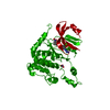
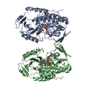
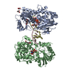
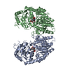
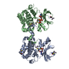
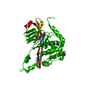
- PDB-1y8z: alpha-glucosyltransferase in complex with UDP and a 13-mer DNA co... -
+ Open data

- Basic information
| Entry | Database: PDB / ID: 1y8z | ||||||
|---|---|---|---|---|---|---|---|
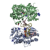
| Title | alpha-glucosyltransferase in complex with UDP and a 13-mer DNA containing a HMU base at 1.9 A resolution | ||||||
 Components Components |
| ||||||
 Keywords Keywords | Transferase/DNA / Transferase / Transferase-DNA COMPLEX | ||||||
| Function / homology |  Function and homology information Function and homology informationDNA alpha-glucosyltransferase / DNA alpha-glucosyltransferase activity / symbiont-mediated suppression of host adaptive immune response / symbiont-mediated suppression of host CRISPR-cas system / symbiont-mediated evasion of host restriction-modification system / DNA modification / symbiont-mediated suppression of host innate immune response Similarity search - Function | ||||||
| Biological species |  Enterobacteria phage T4 (virus) Enterobacteria phage T4 (virus) | ||||||
| Method |  X-RAY DIFFRACTION / SYNCHROTRON / MOLECULAR REPLACEMENT / Resolution: 1.9 Å X-RAY DIFFRACTION / SYNCHROTRON / MOLECULAR REPLACEMENT / Resolution: 1.9 Å | ||||||
 Authors Authors | Lariviere, L. / Sommer, N. / Morera, S. | ||||||
 Citation Citation | Journal: J.Mol.Biol. / Year: 2005 Title: Structural evidence of a passive base-flipping mechanism for AGT, an unusual GT-B glycosyltransferase. Authors: Lariviere, L. / Sommer, N. / Morera, S. | ||||||
| History |
|
- Structure visualization
| Structure viewer | Molecule: MolmilJmol/JSmol |
|---|
- Downloads & links
-Download
| PDBx/mmCIF format | 1y8z.cif.gz | 197.6 KB | Display | PDBx/mmCIF format |
|---|---|---|---|---|
| PDB format | pdb1y8z.ent.gz | 152.1 KB | Display | PDB format |
| PDBx/mmJSON format | 1y8z.json.gz | Tree view | PDBx/mmJSON format | |
| Others |  Other downloads Other downloads |
-Validation report
| Arichive directory | https://data.pdbj.org/pub/pdb/validation_reports/y8/1y8zftp://data.pdbj.org/pub/pdb/validation_reports/y8/1y8z | HTTPS FTP |
|---|
-Related structure data
| Related structure data |  1xv5C 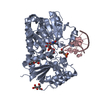 1y6fSC 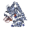 1y6gC 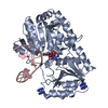 1ya6C C: citing same article ( S: Starting model for refinement |
|---|---|
| Similar structure data |


-Links
 PDBj
PDBj


- Assembly
| Deposited unit | 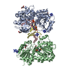
| ||||||||
|---|---|---|---|---|---|---|---|---|---|
| 1 | 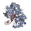
| ||||||||
| Unit cell |
|
-Components
-DNA chain , 2 types, 2 molecules CD
| #1: DNA chain | Mass: 4030.647 Da / Num. of mol.: 1 / Source method: obtained synthetically |
|---|---|
| #2: DNA chain | Mass: 2730.810 Da / Num. of mol.: 1 / Source method: obtained synthetically |
-Protein , 1 types, 2 molecules AB
| #3: Protein | Mass: 47062.426 Da / Num. of mol.: 2 Source method: isolated from a genetically manipulated source Source: (gene. exp.) Enterobacteria phage T4 (virus) / Genus: T4-like viruses / Species: Enterobacteria phage T4 sensu lato / Plasmid: proExHTb / Production host:  |
|---|
-Non-polymers , 5 types, 506 molecules 


| #4: Chemical | ChemComp-NCO /  Mass: 161.116 Da / Num. of mol.: 5 / Source method: obtained synthetically / Formula: CoH18N6 Mass: 161.116 Da / Num. of mol.: 5 / Source method: obtained synthetically / Formula: CoH18N6#5: Chemical |  Type: RNA linking / Mass: 404.161 Da / Num. of mol.: 2 / Source method: obtained synthetically / Formula: C9H14N2O12P2 / Comment: UDP*YM Type: RNA linking / Mass: 404.161 Da / Num. of mol.: 2 / Source method: obtained synthetically / Formula: C9H14N2O12P2 / Comment: UDP*YM#6: Chemical | ChemComp-GOL /  Mass: 92.094 Da / Num. of mol.: 4 / Source method: obtained synthetically / Formula: C3H8O3 Mass: 92.094 Da / Num. of mol.: 4 / Source method: obtained synthetically / Formula: C3H8O3#7: Chemical | ChemComp-CL / |  Mass: 35.453 Da / Num. of mol.: 1 / Source method: obtained synthetically / Formula: Cl Mass: 35.453 Da / Num. of mol.: 1 / Source method: obtained synthetically / Formula: Cl#8: Water | ChemComp-HOH / | Mass: 18.015 Da / Num. of mol.: 494 / Source method: isolated from a natural source / Formula: H2O |
|---|
-Details
| Has protein modification | Y |
|---|
-Experimental details
-Experiment
| Experiment | Method: X-RAY DIFFRACTION / Number of used crystals: 1 |
|---|
- Sample preparation
| Crystal | Density Matthews: 2.1 Å3/Da / Density % sol: 41.6 % | ||||||||||||||||||||
|---|---|---|---|---|---|---|---|---|---|---|---|---|---|---|---|---|---|---|---|---|---|
| Crystal grow | Temperature: 298 K / Method: vapor diffusion, hanging drop / pH: 8.5 Details: PEG10000, pH 8.5, VAPOR DIFFUSION, HANGING DROP, temperature 298K | ||||||||||||||||||||
| Components of the solutions |
|
-Data collection
| Diffraction | Mean temperature: 100 K |
|---|---|
| Diffraction source | Source: SYNCHROTRON / Site: ESRF  / Beamline: ID14-1 / Wavelength: 0.934 Å / Beamline: ID14-1 / Wavelength: 0.934 Å |
| Detector | Type: MARRESEARCH / Detector: CCD / Date: Jul 23, 2004 |
| Radiation | Monochromator: 0.97 / Protocol: SINGLE WAVELENGTH / Monochromatic (M) / Laue (L): M / Scattering type: x-ray |
| Radiation wavelength | Wavelength: 0.934 Å / Relative weight: 1 |
| Reflection | Resolution: 1.9→20 Å / Num. all: 62948 / Num. obs: 62256 / % possible obs: 99 % / Observed criterion σ(F): 2 / Observed criterion σ(I): 1 / Redundancy: 7 % / Biso Wilson estimate: 25 Å2 / Rsym value: 0.058 / Net I/σ(I): 7.8 |
| Reflection shell | Resolution: 1.9→1.97 Å / Mean I/σ(I) obs: 2.2 / Rsym value: 0.45 / % possible all: 93.6 |
- Processing
| Software |
| |||||||||||||||||||||||||
|---|---|---|---|---|---|---|---|---|---|---|---|---|---|---|---|---|---|---|---|---|---|---|---|---|---|---|
| Refinement | Method to determine structure: MOLECULAR REPLACEMENT Starting model: 1Y6F Resolution: 1.9→20 Å / σ(F): 1 / Stereochemistry target values: Engh & Huber
| |||||||||||||||||||||||||
| Refinement step | Cycle: LAST / Resolution: 1.9→20 Å
| |||||||||||||||||||||||||
| Refine LS restraints |
|
