 Movie
Movie Controller

[English] 日本語
 Yorodumi
- PDB-4uc2: Crystal structure of translocator protein 18kDa (TSPO) from rhodo... -
+ Open data

- Basic information
| Entry | Database: PDB / ID: 4uc2 | ||||||
|---|---|---|---|---|---|---|---|
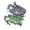
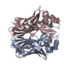
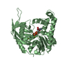
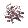
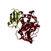
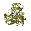
| Title | Crystal structure of translocator protein 18kDa (TSPO) from rhodobacter sphaeroides (A139T mutant) in P212121 space group | ||||||
 Components Components | TRANSLOCATOR PROTEIN TSPO | ||||||
 Keywords Keywords | MEMBRANE PROTEIN / mitochondria / transport / transmembrane protein | ||||||
| Function / homology |  Function and homology information Function and homology informationtetrapyrrole metabolic process / tetrapyrrole binding / lipid binding / membrane / identical protein binding / plasma membrane Similarity search - Function | ||||||
| Biological species |  Rhodobacter sphaeroides (bacteria) Rhodobacter sphaeroides (bacteria) | ||||||
| Method |  X-RAY DIFFRACTION / SYNCHROTRON / MOLECULAR REPLACEMENT / Resolution: 2.4 Å X-RAY DIFFRACTION / SYNCHROTRON / MOLECULAR REPLACEMENT / Resolution: 2.4 Å | ||||||
 Authors Authors | Li, F. / Liu, J. / Zheng, Y. / Garavito, R.M. / Ferguson-Miller, S. | ||||||
| Funding support |  United States, 1items United States, 1items
| ||||||
 Citation Citation | Journal: Science / Year: 2015 Title: Crystal structures of translocator protein (TSPO) and mutant mimic of a human polymorphism. Authors: Li, F. / Liu, J. / Zheng, Y. / Garavito, R.M. / Ferguson-Miller, S. | ||||||
| History |
|
- Structure visualization
| Structure viewer | Molecule: MolmilJmol/JSmol |
|---|
- Downloads & links
-Download
| PDBx/mmCIF format | 4uc2.cif.gz | 79.9 KB | Display | PDBx/mmCIF format |
|---|---|---|---|---|
| PDB format | pdb4uc2.ent.gz | 58.7 KB | Display | PDB format |
| PDBx/mmJSON format | 4uc2.json.gz | Tree view | PDBx/mmJSON format | |
| Others |  Other downloads Other downloads |
-Validation report
| Arichive directory | https://data.pdbj.org/pub/pdb/validation_reports/uc/4uc2ftp://data.pdbj.org/pub/pdb/validation_reports/uc/4uc2 | HTTPS FTP |
|---|
-Related structure data
| Related structure data |  4uc1SC  4uc3C S: Starting model for refinement C: citing same article ( |
|---|---|
| Similar structure data |


-Links
 PDBj
PDBj


- Assembly
| Deposited unit | 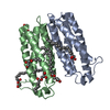
| ||||||||
|---|---|---|---|---|---|---|---|---|---|
| 1 |
| ||||||||
| Unit cell |
|
-Components
| #1: Protein | Mass: 17812.791 Da / Num. of mol.: 2 / Mutation: A139T Source method: isolated from a genetically manipulated source Source: (gene. exp.) Rhodobacter sphaeroides (bacteria) / Production host: #2: Chemical | ChemComp-OLC / (   Mass: 356.540 Da / Num. of mol.: 6 / Source method: obtained synthetically / Formula: C21H40O4 Mass: 356.540 Da / Num. of mol.: 6 / Source method: obtained synthetically / Formula: C21H40O4#3: Chemical | ChemComp-PG4 / |   Mass: 194.226 Da / Num. of mol.: 1 / Source method: obtained synthetically / Formula: C8H18O5 / Comment: precipitant*YM Mass: 194.226 Da / Num. of mol.: 1 / Source method: obtained synthetically / Formula: C8H18O5 / Comment: precipitant*YM#4: Water | ChemComp-HOH / |  Mass: 18.015 Da / Num. of mol.: 35 / Source method: isolated from a natural source / Formula: H2O Mass: 18.015 Da / Num. of mol.: 35 / Source method: isolated from a natural source / Formula: H2O |
|---|
-Experimental details
-Experiment
| Experiment | Method: X-RAY DIFFRACTION / Number of used crystals: 1 |
|---|
- Sample preparation
| Crystal | Density Matthews: 2.78 Å3/Da / Density % sol: 55.78 % |
|---|---|
| Crystal grow | Temperature: 293 K / Method: lipidic cubic phase / pH: 8.5 Details: 23% PEG 3350, 0.1 M (NH4)2SO4 and 0.1 M Hepes buffer pH 8.5 |
-Data collection
| Diffraction | Mean temperature: 100 K |
|---|---|
| Diffraction source | Source: SYNCHROTRON / Site: APS / Beamline: 23-ID-B / Wavelength: 1.003 Å |
| Detector | Type: MARMOSAIC 300 mm CCD / Detector: CCD / Date: Jun 21, 2014 |
| Radiation | Protocol: SINGLE WAVELENGTH / Monochromatic (M) / Laue (L): M / Scattering type: x-ray |
| Radiation wavelength | Wavelength: 1.003 Å / Relative weight: 1 |
| Reflection | Resolution: 2.4→43.65 Å / Num. obs: 29986 / % possible obs: 100 % / Redundancy: 7.1 % / Biso Wilson estimate: 42.36 Å2 / Net I/σ(I): 10.8 |
| Reflection shell | Resolution: 2.4→2.49 Å / Redundancy: 7.3 % / Mean I/σ(I) obs: 2 / % possible all: 100 |
- Processing
| Software |
| ||||||||||||||||||||||||||||||||||||||||||||||||||||||||||||||||||||||
|---|---|---|---|---|---|---|---|---|---|---|---|---|---|---|---|---|---|---|---|---|---|---|---|---|---|---|---|---|---|---|---|---|---|---|---|---|---|---|---|---|---|---|---|---|---|---|---|---|---|---|---|---|---|---|---|---|---|---|---|---|---|---|---|---|---|---|---|---|---|---|---|
| Refinement | Method to determine structure: MOLECULAR REPLACEMENT Starting model: 4UC1 Resolution: 2.4→43.65 Å / SU ML: 0.33 / Cross valid method: FREE R-VALUE / σ(F): 1.34 / Phase error: 24.76 / Stereochemistry target values: ML
| ||||||||||||||||||||||||||||||||||||||||||||||||||||||||||||||||||||||
| Solvent computation | Shrinkage radii: 0.9 Å / VDW probe radii: 1.11 Å / Solvent model: FLAT BULK SOLVENT MODEL | ||||||||||||||||||||||||||||||||||||||||||||||||||||||||||||||||||||||
| Displacement parameters | Biso max: 84.81 Å2 / Biso mean: 42.4165 Å2 / Biso min: 16.06 Å2 | ||||||||||||||||||||||||||||||||||||||||||||||||||||||||||||||||||||||
| Refinement step | Cycle: final / Resolution: 2.4→43.65 Å
| ||||||||||||||||||||||||||||||||||||||||||||||||||||||||||||||||||||||
| Refine LS restraints |
| ||||||||||||||||||||||||||||||||||||||||||||||||||||||||||||||||||||||
| LS refinement shell | Refine-ID: X-RAY DIFFRACTION / Total num. of bins used: 9
|
