 Movie
Movie Controller

[English] 日本語
 Yorodumi
- PDB-4u87: Crystal structure of the Ba-soaked C2 crystal form of pMV158 repl... -
+ Open data

- Basic information
| Entry | Database: PDB / ID: 4u87 | |||||||||||||||
|---|---|---|---|---|---|---|---|---|---|---|---|---|---|---|---|---|
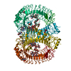
| Title | Crystal structure of the Ba-soaked C2 crystal form of pMV158 replication initiator RepB (P3221 space group) | |||||||||||||||
 Components Components | Replication protein RepB | |||||||||||||||
 Keywords Keywords | REPLICATION / DNA replication initiator | |||||||||||||||
| Function / homology |  Function and homology information Function and homology informationDNA topoisomerase activity / extrachromosomal circular DNA / DNA replication / DNA binding / identical protein binding Similarity search - Function | |||||||||||||||
| Biological species |  Streptococcus agalactiae (bacteria) Streptococcus agalactiae (bacteria) | |||||||||||||||
| Method |  X-RAY DIFFRACTION / SYNCHROTRON / MOLECULAR REPLACEMENT / Resolution: 3.8 Å X-RAY DIFFRACTION / SYNCHROTRON / MOLECULAR REPLACEMENT / Resolution: 3.8 Å | |||||||||||||||
 Authors Authors | Boer, D.R. / Ruiz Maso, J.A. / del Solar, G. / Coll, M. | |||||||||||||||
| Funding support |  Spain, 4items Spain, 4items
| |||||||||||||||
 Citation Citation | Journal: Sci Rep / Year: 2016 Title: Conformational plasticity of RepB, the replication initiator protein of promiscuous streptococcal plasmid pMV158. Authors: Boer, D.R. / Ruiz-Maso, J.A. / Rueda, M. / Petoukhov, M.V. / Machon, C. / Svergun, D.I. / Orozco, M. / Del Solar, G. / Coll, M. | |||||||||||||||
| History |
|
- Structure visualization
| Structure viewer | Molecule: MolmilJmol/JSmol |
|---|
- Downloads & links
-Download
| PDBx/mmCIF format | 4u87.cif.gz | 258.1 KB | Display | PDBx/mmCIF format |
|---|---|---|---|---|
| PDB format | pdb4u87.ent.gz | 211.9 KB | Display | PDB format |
| PDBx/mmJSON format | 4u87.json.gz | Tree view | PDBx/mmJSON format | |
| Others |  Other downloads Other downloads |
-Validation report
| Arichive directory | https://data.pdbj.org/pub/pdb/validation_reports/u8/4u87ftp://data.pdbj.org/pub/pdb/validation_reports/u8/4u87 | HTTPS FTP |
|---|
-Related structure data
| Related structure data |  3dkxS S: Starting model for refinement |
|---|---|
| Similar structure data |


-Links
 PDBj
PDBj- Assembly
| Deposited unit | 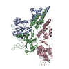
| |||||||||||||||||||||||||||||||||||||||||||||
|---|---|---|---|---|---|---|---|---|---|---|---|---|---|---|---|---|---|---|---|---|---|---|---|---|---|---|---|---|---|---|---|---|---|---|---|---|---|---|---|---|---|---|---|---|---|---|
| 1 | 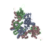
| |||||||||||||||||||||||||||||||||||||||||||||
| Unit cell |
| |||||||||||||||||||||||||||||||||||||||||||||
| Symmetry | Helical symmetry: (Circular symmetry: 2 / Dyad axis: no) | |||||||||||||||||||||||||||||||||||||||||||||
| Noncrystallographic symmetry (NCS) | NCS domain:
NCS domain segments:
NCS ensembles :
|
-Components
| #1: Protein | Mass: 24287.105 Da / Num. of mol.: 3 Source method: isolated from a genetically manipulated source Details: plasmid pMV158 / Source: (gene. exp.) Streptococcus agalactiae (bacteria) / Gene: repB / Plasmid: pGEM-T / Production host: #2: Chemical |   Mass: 35.453 Da / Num. of mol.: 3 / Source method: obtained synthetically / Formula: Cl Mass: 35.453 Da / Num. of mol.: 3 / Source method: obtained synthetically / Formula: Cl#3: Chemical |   Mass: 54.938 Da / Num. of mol.: 3 / Source method: obtained synthetically / Formula: Mn Mass: 54.938 Da / Num. of mol.: 3 / Source method: obtained synthetically / Formula: Mn#4: Chemical | ChemComp-BA /   Mass: 137.327 Da / Num. of mol.: 9 / Source method: obtained synthetically / Formula: Ba Mass: 137.327 Da / Num. of mol.: 9 / Source method: obtained synthetically / Formula: Ba |
|---|
-Experimental details
-Experiment
| Experiment | Method: X-RAY DIFFRACTION |
|---|
- Sample preparation
| Crystal | Density Matthews: 3.59 Å3/Da / Density % sol: 65.74 % |
|---|---|
| Crystal grow | Temperature: 298 K / Method: vapor diffusion, sitting drop / Details: 12% PEG 8000, 100mM BaCl2, 50mM TRIS HCl, pH 8.5 |
-Data collection
| Diffraction | Mean temperature: 100 K |
|---|---|
| Diffraction source | Source: SYNCHROTRON / Site: ESRF  / Beamline: BM16 / Wavelength: 2.0702 Å / Beamline: BM16 / Wavelength: 2.0702 Å |
| Detector | Type: MAR CCD 165 mm / Detector: CCD / Date: Feb 16, 2005 |
| Radiation | Monochromator: double crystal (Si111) monochromator / Protocol: SINGLE WAVELENGTH / Monochromatic (M) / Laue (L): M / Scattering type: x-ray |
| Radiation wavelength | Wavelength: 2.0702 Å / Relative weight: 1 |
| Reflection | Resolution: 3.8→19.77 Å / Num. obs: 19844 / % possible obs: 99.3 % / Observed criterion σ(I): -3 / Redundancy: 7.54 % / Biso Wilson estimate: 67 Å2 / Rmerge(I) obs: 0.236 / Net I/σ(I): 7.54 |
| Reflection shell | Resolution: 3.8→4 Å / Redundancy: 6.9 % / Rmerge(I) obs: 0.731 / Mean I/σ(I) obs: 2.82 / % possible all: 100 |
- Processing
| Software |
| ||||||||||||||||||||||||||||||||||||||||||||||||||||||||||||||||||||||||||||||||||||||||||||||||||||
|---|---|---|---|---|---|---|---|---|---|---|---|---|---|---|---|---|---|---|---|---|---|---|---|---|---|---|---|---|---|---|---|---|---|---|---|---|---|---|---|---|---|---|---|---|---|---|---|---|---|---|---|---|---|---|---|---|---|---|---|---|---|---|---|---|---|---|---|---|---|---|---|---|---|---|---|---|---|---|---|---|---|---|---|---|---|---|---|---|---|---|---|---|---|---|---|---|---|---|---|---|---|
| Refinement | Method to determine structure: MOLECULAR REPLACEMENT Starting model: 3DKX Resolution: 3.8→19.77 Å / SU ML: 0.48 / Cross valid method: FREE R-VALUE / σ(F): 1.37 / Phase error: 24.12 / Stereochemistry target values: ML
| ||||||||||||||||||||||||||||||||||||||||||||||||||||||||||||||||||||||||||||||||||||||||||||||||||||
| Solvent computation | Shrinkage radii: 0.9 Å / VDW probe radii: 1.11 Å / Solvent model: FLAT BULK SOLVENT MODEL / Bsol: 43.59 Å2 / ksol: 0.278 e/Å3 | ||||||||||||||||||||||||||||||||||||||||||||||||||||||||||||||||||||||||||||||||||||||||||||||||||||
| Displacement parameters | Biso mean: 109 Å2
| ||||||||||||||||||||||||||||||||||||||||||||||||||||||||||||||||||||||||||||||||||||||||||||||||||||
| Refinement step | Cycle: LAST / Resolution: 3.8→19.77 Å
| ||||||||||||||||||||||||||||||||||||||||||||||||||||||||||||||||||||||||||||||||||||||||||||||||||||
| Refine LS restraints |
| ||||||||||||||||||||||||||||||||||||||||||||||||||||||||||||||||||||||||||||||||||||||||||||||||||||
| Refine LS restraints NCS |
| ||||||||||||||||||||||||||||||||||||||||||||||||||||||||||||||||||||||||||||||||||||||||||||||||||||
| LS refinement shell |
| ||||||||||||||||||||||||||||||||||||||||||||||||||||||||||||||||||||||||||||||||||||||||||||||||||||
| Refinement TLS params. | Method: refined / Refine-ID: X-RAY DIFFRACTION
| ||||||||||||||||||||||||||||||||||||||||||||||||||||||||||||||||||||||||||||||||||||||||||||||||||||
| Refinement TLS group |
|
