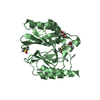
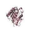
Entry Database : PDB / ID : 4u5xTitle Structure of plant small GTPase OsRac1 complexed with the non-hydrolyzable GTP analog GMPPNP Rac-like GTP-binding protein 1 Keywords / / / Function / homology Function Domain/homology Component
/ / / / / / / / / / / / / / / / / / / / / / / / / / / / / / / / / / / / / / / / / / / / / Biological species Oryza sativa subsp. japonica (Japanese rice)Method / / / Resolution : 1.9 Å Authors Ohki, I. / Kosami, K. / Fujiwara, T. / Nakagawa, A. / Shimamoto, K. / Kojima, C. Journal : J.Biol.Chem. / Year : 2014Title : The Crystal Structure of the Plant Small GTPase OsRac1 Reveals Its Mode of Binding to NADPH OxidaseAuthors : Kosami, K. / Ohki, I. / Nagano, M. / Furuita, K. / Sugiki, T. / Kawano, Y. / Kawasaki, T. / Fujiwara, T. / Nakagawa, A. / Shimamoto, K. / Kojima, C. History Deposition Jul 25, 2014 Deposition site / Processing site Revision 1.0 Aug 20, 2014 Provider / Type Revision 1.1 Oct 22, 2014 Group Revision 1.2 Jan 29, 2020 Group Data collection / Database references ... Data collection / Database references / Derived calculations / Other / Source and taxonomy / Structure summary Category citation / diffrn_source ... citation / diffrn_source / entity_src_gen / pdbx_database_status / pdbx_struct_assembly / pdbx_struct_assembly_prop / pdbx_struct_oper_list / struct_keywords Item _citation.journal_id_CSD / _diffrn_source.pdbx_synchrotron_site ... _citation.journal_id_CSD / _diffrn_source.pdbx_synchrotron_site / _entity_src_gen.pdbx_alt_source_flag / _pdbx_database_status.pdb_format_compatible / _pdbx_struct_assembly.oligomeric_details / _pdbx_struct_assembly_prop.type / _pdbx_struct_assembly_prop.value / _pdbx_struct_oper_list.symmetry_operation / _struct_keywords.text Revision 1.3 Nov 8, 2023 Group Data collection / Database references ... Data collection / Database references / Derived calculations / Refinement description Category chem_comp_atom / chem_comp_bond ... chem_comp_atom / chem_comp_bond / database_2 / pdbx_initial_refinement_model / pdbx_struct_conn_angle / refine_hist / struct_conn Item _database_2.pdbx_DOI / _database_2.pdbx_database_accession ... _database_2.pdbx_DOI / _database_2.pdbx_database_accession / _pdbx_struct_conn_angle.ptnr1_auth_seq_id / _pdbx_struct_conn_angle.ptnr3_auth_seq_id / _pdbx_struct_conn_angle.value / _refine_hist.number_atoms_solvent / _refine_hist.pdbx_number_atoms_ligand / _refine_hist.pdbx_number_atoms_nucleic_acid / _refine_hist.pdbx_number_atoms_protein / _struct_conn.pdbx_dist_value / _struct_conn.ptnr2_auth_seq_id
Show all Show less
 Movie
Movie Controller

 Yorodumi
 Open data
 Basic information
 Components
 Keywords
 Function and homology information
 X-RAY DIFFRACTION /
  Authors
 Citation
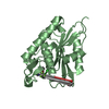
 Structure visualization
 Downloads & links
 Other downloads
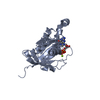


 PDBj
PDBj


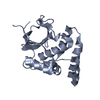
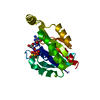
 Assembly
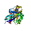


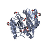
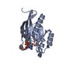
 Mass: 522.196 Da / Num. of mol.: 1 / Source method: obtained synthetically / Formula: C10H17N6O13P3
 Mass: 24.305 Da / Num. of mol.: 1 / Source method: obtained synthetically / Formula: Mg
 Mass: 92.094 Da / Num. of mol.: 3 / Source method: obtained synthetically / Formula: C3H8O3
 Mass: 18.015 Da / Num. of mol.: 90 / Source method: isolated from a natural source / Formula: H2O
 Sample preparation
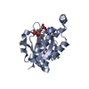
 / Beamline: BL44XU / Wavelength: 0.9 Å
 Processing