 Movie
Movie Controller

[English] 日本語
 Yorodumi
- PDB-4tyb: An Ligand-observed Mass Spectrometry-based Approach Integrated in... -
+ Open data

- Basic information
| Entry | Database: PDB / ID: 4tyb | ||||||
|---|---|---|---|---|---|---|---|
| Title | An Ligand-observed Mass Spectrometry-based Approach Integrated into the Fragment Based Lead Discovery Pipeline | ||||||
 Components Components | Polyprotein | ||||||
 Keywords Keywords | TRANSFERASE/TRANSFERASE INHIBITOR / Inhibitor / NS5B / TRANSFERASE-TRANSFERASE INHIBITOR complex | ||||||
| Function / homology |  Function and homology information Function and homology informationserine-type peptidase activity / RNA helicase activity / RNA-directed RNA polymerase / viral RNA genome replication / RNA-directed RNA polymerase activity / ATP hydrolysis activity / proteolysis / RNA binding / ATP binding Similarity search - Function | ||||||
| Biological species |  Hepatitis C virus Hepatitis C virus | ||||||
| Method |  X-RAY DIFFRACTION / Resolution: 2.93 Å X-RAY DIFFRACTION / Resolution: 2.93 Å | ||||||
 Authors Authors | Shui, W. / Yang, C. / Lin, J. / Chen, X. / Qin, S. / Chen, S. | ||||||
 Citation Citation | Journal: Sci Rep / Year: 2015 Title: A ligand-observed mass spectrometry approach integrated into the fragment based lead discovery pipeline Authors: Chen, X. / Qin, S. / Chen, S. / Li, J. / Li, L. / Wang, Z. / Wang, Q. / Lin, J. / Yang, C. / Shui, W. | ||||||
| History |
|
- Structure visualization
| Structure viewer | Molecule: MolmilJmol/JSmol |
|---|
- Downloads & links
-Download
| PDBx/mmCIF format | 4tyb.cif.gz | 413.2 KB | Display | PDBx/mmCIF format |
|---|---|---|---|---|
| PDB format | pdb4tyb.ent.gz | 339.8 KB | Display | PDB format |
| PDBx/mmJSON format | 4tyb.json.gz | Tree view | PDBx/mmJSON format | |
| Others |  Other downloads Other downloads |
-Validation report
| Arichive directory | https://data.pdbj.org/pub/pdb/validation_reports/ty/4tybftp://data.pdbj.org/pub/pdb/validation_reports/ty/4tyb | HTTPS FTP |
|---|
-Related structure data
| Related structure data |  4txsC  4ty8C  4ty9C 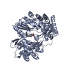 4tyaC  4txf C: citing same article ( |
|---|---|
| Similar structure data |

-Links
 PDBj
PDBj
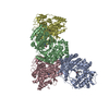
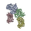
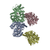
- Assembly
| Deposited unit | 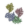
| ||||||||
|---|---|---|---|---|---|---|---|---|---|
| 1 |
| ||||||||
| Unit cell |
|
-Components
| #1: Protein | Mass: 62791.984 Da / Num. of mol.: 4 Source method: isolated from a genetically manipulated source Source: (gene. exp.) Hepatitis C virus / Production host:  #2: Chemical | ChemComp-3B1 / (   Mass: 202.252 Da / Num. of mol.: 4 / Source method: obtained synthetically / Formula: C12H14N2O Mass: 202.252 Da / Num. of mol.: 4 / Source method: obtained synthetically / Formula: C12H14N2O |
|---|
-Experimental details
-Experiment
| Experiment | Method: X-RAY DIFFRACTION |
|---|
- Sample preparation
| Crystal | Density Matthews: 2.61 Å3/Da / Density % sol: 52.92 % |
|---|---|
| Crystal grow | Temperature: 289 K / Method: vapor diffusion, hanging drop Details: PEG4000, Dithiothreitol, 2-(N-morpholino)ethanesulfonic acid, Glycerin PH range: 5.5-6.5 |
-Data collection
| Diffraction | Mean temperature: 100 K |
|---|---|
| Diffraction source | Source: ROTATING ANODE / Type: RIGAKU MICROMAX-007 HF / Wavelength: 1.5415 Å |
| Detector | Type: RIGAKU RAXIS HTC / Detector: IMAGE PLATE / Date: May 23, 2012 |
| Radiation | Monochromator: Cu filter / Protocol: LAUE / Monochromatic (M) / Laue (L): M / Scattering type: x-ray |
| Radiation wavelength | Wavelength: 1.5415 Å / Relative weight: 1 |
| Reflection | Resolution: 2.93→50 Å / Num. obs: 57886 / % possible obs: 99.2 % / Redundancy: 4.9 % / Net I/σ(I): 14.8 |
- Processing
| Software |
| ||||||||||||||||||||||||||||||||||||||||||||||||||||||||||||||||||||||||||||||||||||||||||||||||||||||||||||||||||||||||||||||||||||||||||||||||||||||||||||||||||||||||||||||||||||||
|---|---|---|---|---|---|---|---|---|---|---|---|---|---|---|---|---|---|---|---|---|---|---|---|---|---|---|---|---|---|---|---|---|---|---|---|---|---|---|---|---|---|---|---|---|---|---|---|---|---|---|---|---|---|---|---|---|---|---|---|---|---|---|---|---|---|---|---|---|---|---|---|---|---|---|---|---|---|---|---|---|---|---|---|---|---|---|---|---|---|---|---|---|---|---|---|---|---|---|---|---|---|---|---|---|---|---|---|---|---|---|---|---|---|---|---|---|---|---|---|---|---|---|---|---|---|---|---|---|---|---|---|---|---|---|---|---|---|---|---|---|---|---|---|---|---|---|---|---|---|---|---|---|---|---|---|---|---|---|---|---|---|---|---|---|---|---|---|---|---|---|---|---|---|---|---|---|---|---|---|---|---|---|---|
| Refinement | Resolution: 2.93→47.41 Å / Cor.coef. Fo:Fc: 0.914 / Cor.coef. Fo:Fc free: 0.848 / SU B: 18.05 / SU ML: 0.342 / Cross valid method: THROUGHOUT / ESU R Free: 0.456 / Stereochemistry target values: MAXIMUM LIKELIHOOD / Details: HYDROGENS HAVE BEEN ADDED IN THE RIDING POSITIONS
| ||||||||||||||||||||||||||||||||||||||||||||||||||||||||||||||||||||||||||||||||||||||||||||||||||||||||||||||||||||||||||||||||||||||||||||||||||||||||||||||||||||||||||||||||||||||
| Solvent computation | Ion probe radii: 0.8 Å / Shrinkage radii: 0.8 Å / VDW probe radii: 1.2 Å / Solvent model: MASK | ||||||||||||||||||||||||||||||||||||||||||||||||||||||||||||||||||||||||||||||||||||||||||||||||||||||||||||||||||||||||||||||||||||||||||||||||||||||||||||||||||||||||||||||||||||||
| Displacement parameters | Biso mean: 28.21 Å2
| ||||||||||||||||||||||||||||||||||||||||||||||||||||||||||||||||||||||||||||||||||||||||||||||||||||||||||||||||||||||||||||||||||||||||||||||||||||||||||||||||||||||||||||||||||||||
| Refinement step | Cycle: 1 / Resolution: 2.93→47.41 Å
| ||||||||||||||||||||||||||||||||||||||||||||||||||||||||||||||||||||||||||||||||||||||||||||||||||||||||||||||||||||||||||||||||||||||||||||||||||||||||||||||||||||||||||||||||||||||
| Refine LS restraints |
|