 Movie
Movie Controller

[English] 日本語
 Yorodumi
- PDB-4tsh: A Novel Protein Fold Forms an Intramolecular Lock to Stabilize th... -
+ Open data

- Basic information
| Entry | Database: PDB / ID: 4tsh | ||||||||||||
|---|---|---|---|---|---|---|---|---|---|---|---|---|---|
| Title | A Novel Protein Fold Forms an Intramolecular Lock to Stabilize the Tertiary Structure of Streptococcus mutans Adhesin P1 | ||||||||||||
 Components Components | (Surface protein adhesin) x 2 | ||||||||||||
 Keywords Keywords | CELL ADHESION / Adhesin / Streptococcus / Intramolecular Lock / Complex | ||||||||||||
| Function / homology |  Function and homology information Function and homology informationprotein transport to vacuole involved in ubiquitin-dependent protein catabolic process via the multivesicular body sorting pathway / metal ion binding / identical protein binding Similarity search - Function | ||||||||||||
| Biological species |  Streptococcus mutans (bacteria) Streptococcus mutans (bacteria) | ||||||||||||
| Method |  X-RAY DIFFRACTION / SYNCHROTRON / MOLECULAR REPLACEMENT / Resolution: 2 Å X-RAY DIFFRACTION / SYNCHROTRON / MOLECULAR REPLACEMENT / Resolution: 2 Å | ||||||||||||
 Authors Authors | Heim, K.P. / Kailasan, S. / McKenna, R. / Brady, L.J. | ||||||||||||
| Funding support |  United States, 3items United States, 3items
| ||||||||||||
 Citation Citation | Journal: Proc.Natl.Acad.Sci.USA / Year: 2014 Title: An intramolecular lock facilitates folding and stabilizes the tertiary structure of Streptococcus mutans adhesin P1. Authors: Heim, K.P. / Crowley, P.J. / Long, J.R. / Kailasan, S. / McKenna, R. / Brady, L.J. | ||||||||||||
| History |
|
- Structure visualization
| Structure viewer | Molecule: MolmilJmol/JSmol |
|---|
- Downloads & links
-Download
| PDBx/mmCIF format | 4tsh.cif.gz | 365.2 KB | Display | PDBx/mmCIF format |
|---|---|---|---|---|
| PDB format | pdb4tsh.ent.gz | 300.6 KB | Display | PDB format |
| PDBx/mmJSON format | 4tsh.json.gz | Tree view | PDBx/mmJSON format | |
| Others |  Other downloads Other downloads |
-Validation report
| Arichive directory | https://data.pdbj.org/pub/pdb/validation_reports/ts/4tshftp://data.pdbj.org/pub/pdb/validation_reports/ts/4tsh | HTTPS FTP |
|---|
-Related structure data
| Related structure data | 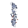 3qe5S S: Starting model for refinement |
|---|---|
| Similar structure data |


-Links
 PDBj
PDBj- Assembly
| Deposited unit | 
| ||||||||
|---|---|---|---|---|---|---|---|---|---|
| 1 |
| ||||||||
| Unit cell |
| ||||||||
| Components on special symmetry positions |
|
-Components
| #1: Protein | Mass: 12813.826 Da / Num. of mol.: 1 / Fragment: UNP residues 52-172 Source method: isolated from a genetically manipulated source Source: (gene. exp.) Streptococcus mutans (bacteria) / Strain: NG8 / Gene: spaP / Production host: | ||||||
|---|---|---|---|---|---|---|---|
| #2: Protein | Mass: 56187.180 Da / Num. of mol.: 1 / Fragment: UNP residues 980-1486 Source method: isolated from a genetically manipulated source Source: (gene. exp.) Streptococcus mutans (bacteria) / Strain: NG8 / Gene: spaP / Production host: | ||||||
| #3: Chemical |   Mass: 40.078 Da / Num. of mol.: 2 / Source method: obtained synthetically / Formula: Ca Mass: 40.078 Da / Num. of mol.: 2 / Source method: obtained synthetically / Formula: Ca#4: Chemical |   Mass: 24.305 Da / Num. of mol.: 2 / Source method: obtained synthetically / Formula: Mg Mass: 24.305 Da / Num. of mol.: 2 / Source method: obtained synthetically / Formula: Mg#5: Water | ChemComp-HOH / |  Mass: 18.015 Da / Num. of mol.: 537 / Source method: isolated from a natural source / Formula: H2O Mass: 18.015 Da / Num. of mol.: 537 / Source method: isolated from a natural source / Formula: H2OHas protein modification | Y | |
-Experimental details
-Experiment
| Experiment | Method: X-RAY DIFFRACTION / Number of used crystals: 1 |
|---|
- Sample preparation
| Crystal | Density Matthews: 4.07 Å3/Da / Density % sol: 69.8 % |
|---|---|
| Crystal grow | Temperature: 295 K / Method: vapor diffusion, hanging drop / pH: 7.5 Details: Crystals were routinely grown at room temperature using the hanging drop vapor diffusion method by mixing 4uL of concentrated NA1/P3C with a mother liquor solution of 24% polyethylene glycol ...Details: Crystals were routinely grown at room temperature using the hanging drop vapor diffusion method by mixing 4uL of concentrated NA1/P3C with a mother liquor solution of 24% polyethylene glycol (PEG) 4000, 150mM ammonium phosphate, at pH 7.5. Crystals formed as large plate clusters within approximately 2 weeks. Following initial crystal growth, 8uL of a solution containing 45% PEG 4000, 100mM ammonium phosphate, at pH 7.5 was added to the crystal drop to facilitate the removal of water from the crystal and improve diffraction quality by producing a more compact and ordered crystal lattice PH range: 7.5 / Temp details: Room Temp |
-Data collection
| Diffraction | Mean temperature: 93 K |
|---|---|
| Diffraction source | Source: SYNCHROTRON / Site: CHESS / Beamline: A1 / Wavelength: 0.987 Å |
| Detector | Type: ADSC QUANTUM 210 / Detector: CCD / Date: May 5, 2014 |
| Radiation | Protocol: SINGLE WAVELENGTH / Monochromatic (M) / Laue (L): M / Scattering type: x-ray |
| Radiation wavelength | Wavelength: 0.987 Å / Relative weight: 1 |
| Reflection | Resolution: 2→35.85 Å / Num. obs: 73061 / % possible obs: 99.8 % / Redundancy: 3.8 % / Rmerge(I) obs: 0.09 / Net I/σ(I): 18.5 |
| Reflection shell | Redundancy: 3.7 % / Rmerge(I) obs: 0.649 / Mean I/σ(I) obs: 2.07 / % possible all: 97 |
- Processing
| Software | Name: PHENIX / Version: (phenix.refine: 1.9_1692) / Classification: refinement | |||||||||||||||||||||||||||||||||||||||||||||||||||||||||||||||||||||||||||||||||||||||||||||||||||||||||||||||||||||||||||||||||||||||||||||||||||||||||||||||||||||||||||||||||||||||||||||||||||||||||||||||||||||||||||||||||||||||||||||||||||||||||||||||||||||||||||||||||||||||||||||||||||||||||||||||||||||||||||||||||||||
|---|---|---|---|---|---|---|---|---|---|---|---|---|---|---|---|---|---|---|---|---|---|---|---|---|---|---|---|---|---|---|---|---|---|---|---|---|---|---|---|---|---|---|---|---|---|---|---|---|---|---|---|---|---|---|---|---|---|---|---|---|---|---|---|---|---|---|---|---|---|---|---|---|---|---|---|---|---|---|---|---|---|---|---|---|---|---|---|---|---|---|---|---|---|---|---|---|---|---|---|---|---|---|---|---|---|---|---|---|---|---|---|---|---|---|---|---|---|---|---|---|---|---|---|---|---|---|---|---|---|---|---|---|---|---|---|---|---|---|---|---|---|---|---|---|---|---|---|---|---|---|---|---|---|---|---|---|---|---|---|---|---|---|---|---|---|---|---|---|---|---|---|---|---|---|---|---|---|---|---|---|---|---|---|---|---|---|---|---|---|---|---|---|---|---|---|---|---|---|---|---|---|---|---|---|---|---|---|---|---|---|---|---|---|---|---|---|---|---|---|---|---|---|---|---|---|---|---|---|---|---|---|---|---|---|---|---|---|---|---|---|---|---|---|---|---|---|---|---|---|---|---|---|---|---|---|---|---|---|---|---|---|---|---|---|---|---|---|---|---|---|---|---|---|---|---|---|---|---|---|---|---|---|---|---|---|---|---|---|---|---|---|---|---|---|---|---|---|---|---|---|---|---|---|---|---|---|---|---|---|---|---|---|---|---|---|---|---|---|---|---|---|---|---|---|---|---|
| Refinement | Method to determine structure: MOLECULAR REPLACEMENT Starting model: 3QE5 Resolution: 2→35.847 Å / SU ML: 0.2 / Cross valid method: THROUGHOUT / σ(F): 1.34 / Phase error: 20.98 / Stereochemistry target values: ML
| |||||||||||||||||||||||||||||||||||||||||||||||||||||||||||||||||||||||||||||||||||||||||||||||||||||||||||||||||||||||||||||||||||||||||||||||||||||||||||||||||||||||||||||||||||||||||||||||||||||||||||||||||||||||||||||||||||||||||||||||||||||||||||||||||||||||||||||||||||||||||||||||||||||||||||||||||||||||||||||||||||||
| Solvent computation | Shrinkage radii: 0.9 Å / VDW probe radii: 1.11 Å / Solvent model: FLAT BULK SOLVENT MODEL | |||||||||||||||||||||||||||||||||||||||||||||||||||||||||||||||||||||||||||||||||||||||||||||||||||||||||||||||||||||||||||||||||||||||||||||||||||||||||||||||||||||||||||||||||||||||||||||||||||||||||||||||||||||||||||||||||||||||||||||||||||||||||||||||||||||||||||||||||||||||||||||||||||||||||||||||||||||||||||||||||||||
| Displacement parameters | Biso max: 280.67 Å2 / Biso mean: 49.0905 Å2 / Biso min: 17.69 Å2 | |||||||||||||||||||||||||||||||||||||||||||||||||||||||||||||||||||||||||||||||||||||||||||||||||||||||||||||||||||||||||||||||||||||||||||||||||||||||||||||||||||||||||||||||||||||||||||||||||||||||||||||||||||||||||||||||||||||||||||||||||||||||||||||||||||||||||||||||||||||||||||||||||||||||||||||||||||||||||||||||||||||
| Refinement step | Cycle: final / Resolution: 2→35.847 Å
| |||||||||||||||||||||||||||||||||||||||||||||||||||||||||||||||||||||||||||||||||||||||||||||||||||||||||||||||||||||||||||||||||||||||||||||||||||||||||||||||||||||||||||||||||||||||||||||||||||||||||||||||||||||||||||||||||||||||||||||||||||||||||||||||||||||||||||||||||||||||||||||||||||||||||||||||||||||||||||||||||||||
| Refine LS restraints |
| |||||||||||||||||||||||||||||||||||||||||||||||||||||||||||||||||||||||||||||||||||||||||||||||||||||||||||||||||||||||||||||||||||||||||||||||||||||||||||||||||||||||||||||||||||||||||||||||||||||||||||||||||||||||||||||||||||||||||||||||||||||||||||||||||||||||||||||||||||||||||||||||||||||||||||||||||||||||||||||||||||||
| LS refinement shell | Refine-ID: X-RAY DIFFRACTION / Total num. of bins used: 14
| |||||||||||||||||||||||||||||||||||||||||||||||||||||||||||||||||||||||||||||||||||||||||||||||||||||||||||||||||||||||||||||||||||||||||||||||||||||||||||||||||||||||||||||||||||||||||||||||||||||||||||||||||||||||||||||||||||||||||||||||||||||||||||||||||||||||||||||||||||||||||||||||||||||||||||||||||||||||||||||||||||||
| Refinement TLS params. | Method: refined / Refine-ID: X-RAY DIFFRACTION
| |||||||||||||||||||||||||||||||||||||||||||||||||||||||||||||||||||||||||||||||||||||||||||||||||||||||||||||||||||||||||||||||||||||||||||||||||||||||||||||||||||||||||||||||||||||||||||||||||||||||||||||||||||||||||||||||||||||||||||||||||||||||||||||||||||||||||||||||||||||||||||||||||||||||||||||||||||||||||||||||||||||
| Refinement TLS group |
|
