 Movie
Movie Controller

[English] 日本語
 Yorodumi
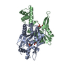
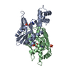
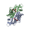
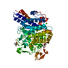
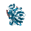
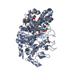
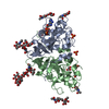
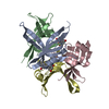
- PDB-4trc: Sulfolobus solfataricus adenine phosphoribosyltransferase with adenine -
+ Open data

- Basic information
| Entry | Database: PDB / ID: 4trc | ||||||
|---|---|---|---|---|---|---|---|
| Title | Sulfolobus solfataricus adenine phosphoribosyltransferase with adenine | ||||||
 Components Components | Purine phosphoribosyltransferase (GpT-1) | ||||||
 Keywords Keywords | TRANSFERASE / Sulfolobus / solfataricus / adenine / phosphoribosyltransferase | ||||||
| Function / homology | Phosphoribosyl transferase domain / Phosphoribosyltransferase-like / Phosphoribosyltransferase domain / glycosyltransferase activity / Transferases; Glycosyltransferases; Pentosyltransferases / nucleotide binding / ADENINE / PHOSPHATE ION / Purine phosphoribosyltransferase (GpT-1) Function and homology information Function and homology information | ||||||
| Biological species |   Sulfolobus solfataricus (archaea) Sulfolobus solfataricus (archaea) | ||||||
| Method |  X-RAY DIFFRACTION / SYNCHROTRON / Resolution: 2.4 Å X-RAY DIFFRACTION / SYNCHROTRON / Resolution: 2.4 Å | ||||||
 Authors Authors | Kadziola, A. | ||||||
 Citation Citation | Journal: Biochemistry / Year: 2015 Title: Adenine Phosphoribosyltransferase from Sulfolobus solfataricus Is an Enzyme with Unusual Kinetic Properties and a Crystal Structure that Suggests It Evolved from a 6-Oxopurine Phosphoribosyltransferase. Authors: Jensen, K.F. / Hansen, M.R. / Jensen, K.S. / Christoffersen, S. / Poulsen, J.C. / Mlgaard, A. / Kadziola, A. | ||||||
| History |
|
- Structure visualization
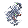
| Structure viewer | Molecule: MolmilJmol/JSmol |
|---|
- Downloads & links
-Download
| PDBx/mmCIF format | 4trc.cif.gz | 100 KB | Display | PDBx/mmCIF format |
|---|---|---|---|---|
| PDB format | pdb4trc.ent.gz | 77.7 KB | Display | PDB format |
| PDBx/mmJSON format | 4trc.json.gz | Tree view | PDBx/mmJSON format | |
| Others |  Other downloads Other downloads |
-Validation report
| Arichive directory | https://data.pdbj.org/pub/pdb/validation_reports/tr/4trcftp://data.pdbj.org/pub/pdb/validation_reports/tr/4trc | HTTPS FTP |
|---|
-Related structure data


-Links
 PDBj
PDBj


- Assembly
| Deposited unit | 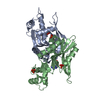
| ||||||||||||||||||
|---|---|---|---|---|---|---|---|---|---|---|---|---|---|---|---|---|---|---|---|
| 1 |
| ||||||||||||||||||
| Unit cell |
| ||||||||||||||||||
| Noncrystallographic symmetry (NCS) | NCS domain:
NCS domain segments:
|
-Components
| #1: Protein | Mass: 24328.211 Da / Num. of mol.: 2 Source method: isolated from a genetically manipulated source Source: (gene. exp.) Sulfolobus solfataricus (archaea) / Strain: ATCC 35092 / DSM 1617 / JCM 11322 / P2 / Gene: gpT-1, SSO2342 / Production host:  References: UniProt: Q97W95, Transferases; Glycosyltransferases; Pentosyltransferases #2: Chemical |   Mass: 135.127 Da / Num. of mol.: 2 / Source method: obtained synthetically / Formula: C5H5N5 Mass: 135.127 Da / Num. of mol.: 2 / Source method: obtained synthetically / Formula: C5H5N5#3: Chemical | ChemComp-PO4 /   Mass: 94.971 Da / Num. of mol.: 5 / Source method: obtained synthetically / Formula: PO4 Mass: 94.971 Da / Num. of mol.: 5 / Source method: obtained synthetically / Formula: PO4#4: Water | ChemComp-HOH / |  Mass: 18.015 Da / Num. of mol.: 157 / Source method: isolated from a natural source / Formula: H2O Mass: 18.015 Da / Num. of mol.: 157 / Source method: isolated from a natural source / Formula: H2O |
|---|
-Experimental details
-Experiment
| Experiment | Method: X-RAY DIFFRACTION |
|---|
- Sample preparation
| Crystal | Density Matthews: 3 Å3/Da / Density % sol: 59.06 % |
|---|---|
| Crystal grow | Temperature: 293 K / Method: vapor diffusion, sitting drop Details: Protein 8.9 mg/mL 25 mM TRIS pH 7.6 0.1 mM EDTA 5 mM adenine Buffer 0.1 M phosphate-citrate pH 4.2 Precipitant 20 % PEG1000 0.2 M Li2SO4 |
-Data collection
| Diffraction | Mean temperature: 122 K |
|---|---|
| Diffraction source | Source: SYNCHROTRON / Site: MAX II  / Beamline: I911-2 / Wavelength: 1.03911 Å / Beamline: I911-2 / Wavelength: 1.03911 Å |
| Detector | Type: MARRESEARCH / Detector: AREA DETECTOR / Date: Apr 1, 2011 |
| Radiation | Protocol: SINGLE WAVELENGTH / Monochromatic (M) / Laue (L): M / Scattering type: x-ray |
| Radiation wavelength | Wavelength: 1.03911 Å / Relative weight: 1 |
| Reflection | Resolution: 2.4→30 Å / Num. obs: 22808 / % possible obs: 99.6 % / Redundancy: 13.6 % / Net I/σ(I): 28.26 |
- Processing
| Software | Name: PHENIX / Version: (phenix.refine: 1.5_2) / Classification: refinement | |||||||||||||||||||||||||||||||||||||||||||||||||||||||||||||||
|---|---|---|---|---|---|---|---|---|---|---|---|---|---|---|---|---|---|---|---|---|---|---|---|---|---|---|---|---|---|---|---|---|---|---|---|---|---|---|---|---|---|---|---|---|---|---|---|---|---|---|---|---|---|---|---|---|---|---|---|---|---|---|---|---|
| Refinement | Resolution: 2.4→29.705 Å / SU ML: 0.35 / Cross valid method: FREE R-VALUE / σ(F): 1.36 / Phase error: 24.14 / Stereochemistry target values: ML
| |||||||||||||||||||||||||||||||||||||||||||||||||||||||||||||||
| Solvent computation | Shrinkage radii: 0.9 Å / VDW probe radii: 1.11 Å / Solvent model: FLAT BULK SOLVENT MODEL / Bsol: 43.456 Å2 / ksol: 0.351 e/Å3 | |||||||||||||||||||||||||||||||||||||||||||||||||||||||||||||||
| Displacement parameters |
| |||||||||||||||||||||||||||||||||||||||||||||||||||||||||||||||
| Refinement step | Cycle: LAST / Resolution: 2.4→29.705 Å
| |||||||||||||||||||||||||||||||||||||||||||||||||||||||||||||||
| Refine LS restraints |
| |||||||||||||||||||||||||||||||||||||||||||||||||||||||||||||||
| Refine LS restraints NCS |
| |||||||||||||||||||||||||||||||||||||||||||||||||||||||||||||||
| LS refinement shell |
|
