| Entry | Database: PDB / ID: 4tkj
|
|---|
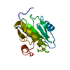
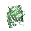
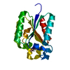
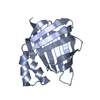
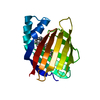
| Title | The 0.87 angstrom X-ray structure of the human heart fatty acid-binding protein complexed with palmitic acid |
|---|
 Components Components | Fatty acid-binding protein, heart |
|---|
 Keywords Keywords | LIPID BINDING PROTEIN / antiparallel beta barrel / fatty acid-binding protein / human heart / palmitic acid |
|---|
| Function / homology |  Function and homology information Function and homology information
icosatetraenoic acid binding / positive regulation of long-chain fatty acid import into cell / regulation of phosphatidylcholine biosynthetic process / regulation of fatty acid oxidation / oleic acid binding / positive regulation of phospholipid biosynthetic process / intracellular lipid transport / response to fatty acid / phospholipid homeostasis / long-chain fatty acid transmembrane transporter activity ...icosatetraenoic acid binding / positive regulation of long-chain fatty acid import into cell / regulation of phosphatidylcholine biosynthetic process / regulation of fatty acid oxidation / oleic acid binding / positive regulation of phospholipid biosynthetic process / intracellular lipid transport / response to fatty acid / phospholipid homeostasis / long-chain fatty acid transmembrane transporter activity / long-chain fatty acid binding / Triglyceride catabolism / sarcoplasm / long-chain fatty acid transport / cytoskeletal protein binding / brown fat cell differentiation / cholesterol homeostasis / fatty acid metabolic process / response to insulin / response to xenobiotic stimulus / negative regulation of cell population proliferation / : / extracellular exosome / nucleus / cytosolSimilarity search - Function Cytosolic fatty-acid binding proteins signature. / Intracellular lipid binding protein / Cytosolic fatty-acid binding / Lipocalin / cytosolic fatty-acid binding protein family / Lipocalin/cytosolic fatty-acid binding domain / Calycin beta-barrel core domain / Calycin / Lipocalin / Beta Barrel / Mainly BetaSimilarity search - Domain/homology |
|---|
| Biological species |  Homo sapiens (human) Homo sapiens (human) |
|---|
| Method |  X-RAY DIFFRACTION / SYNCHROTRON / FOURIER SYNTHESIS / Resolution: 0.87 Å X-RAY DIFFRACTION / SYNCHROTRON / FOURIER SYNTHESIS / Resolution: 0.87 Å |
|---|
 Authors Authors | Sugiyama, S. / Matsuoka, S. / Mizohata, E. / Matsuoka, D. / Ishida, H. / Hirose, M. / Kakinouchi, K. / Hara, T. / Murakami, S. / Inoue, T. / Murata, M. |
|---|
| Funding support |  Japan, 3items Japan, 3items | Organization | Grant number | Country |
|---|
| JST ERATO | 1859 | Japan | | JSPS KAKENHI | 25286051 | Japan | | JSPS KAKENHI | 25650051 | Japan |
|
|---|
 Citation Citation | Journal: Angew.Chem.Int.Ed.Engl. / Year: 2015
Title: Water-mediated recognition of simple alkyl chains by heart-type Fatty-Acid-binding protein
Authors: Matsuoka, S. / Sugiyama, S. / Matsuoka, D. / Hirose, M. / Lethu, S. / Ano, H. / Hara, T. / Ichihara, O. / Kimura, S.R. / Murakami, S. / Ishida, H. / Mizohata, E. / Inoue, T. / Murata, M. |
|---|
| History | | Deposition | May 26, 2014 | Deposition site: RCSB / Processing site: PDBJ |
|---|
| Revision 1.0 | Jan 28, 2015 | Provider: repository / Type: Initial release |
|---|
| Revision 1.1 | Feb 4, 2015 | Group: Database references |
|---|
| Revision 1.2 | Jan 29, 2020 | Group: Advisory / Data collection ...Advisory / Data collection / Database references / Derived calculations / Other / Source and taxonomy / Structure summary
Category: citation / diffrn_source ...citation / diffrn_source / entity / entity_src_gen / pdbx_database_status / pdbx_entity_nonpoly / pdbx_struct_assembly / pdbx_struct_assembly_prop / pdbx_struct_oper_list / pdbx_validate_close_contact
Item: _citation.journal_id_CSD / _diffrn_source.pdbx_synchrotron_site ..._citation.journal_id_CSD / _diffrn_source.pdbx_synchrotron_site / _entity.pdbx_description / _entity.src_method / _entity_src_gen.pdbx_alt_source_flag / _pdbx_database_status.pdb_format_compatible / _pdbx_entity_nonpoly.name / _pdbx_struct_assembly.oligomeric_details / _pdbx_struct_assembly_prop.type / _pdbx_struct_assembly_prop.value / _pdbx_struct_oper_list.symmetry_operation |
|---|
| Revision 1.3 | Jul 29, 2020 | Group: Data collection / Derived calculations ...Data collection / Derived calculations / Refinement description / Structure summary
Category: chem_comp / entity ...chem_comp / entity / pdbx_chem_comp_identifier / pdbx_entity_nonpoly / refine_hist / struct_site / struct_site_gen
Item: _chem_comp.mon_nstd_flag / _chem_comp.name ..._chem_comp.mon_nstd_flag / _chem_comp.name / _chem_comp.type / _entity.pdbx_description / _pdbx_entity_nonpoly.name / _refine_hist.number_atoms_solvent / _refine_hist.number_atoms_total / _refine_hist.pdbx_number_atoms_ligand / _refine_hist.pdbx_number_atoms_nucleic_acid / _refine_hist.pdbx_number_atoms_protein
Description: Carbohydrate remediation / Provider: repository / Type: Remediation |
|---|
| Revision 1.4 | Mar 20, 2024 | Group: Data collection / Database references / Structure summary
Category: chem_comp / chem_comp_atom ...chem_comp / chem_comp_atom / chem_comp_bond / database_2
Item: _chem_comp.pdbx_synonyms / _database_2.pdbx_DOI / _database_2.pdbx_database_accession |
|---|
|
|---|
 Movie
Movie Controller

 Yorodumi
 Open data
 Basic information
 Structure visualization
 Downloads & links
 Other downloads
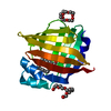


 PDBj
PDBj


 Assembly


 Mass: 256.424 Da / Num. of mol.: 1 / Source method: obtained synthetically / Formula: C16H32O2
 Mass: 282.331 Da / Num. of mol.: 2 / Source method: obtained synthetically / Formula: C12H26O7 / Comment: precipitant*YM
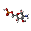
 Type: D-saccharide, alpha linking / Mass: 259.151 Da / Num. of mol.: 1
 Mass: 18.015 Da / Num. of mol.: 189 / Source method: isolated from a natural source / Formula: H2O
 Sample preparation
 Processing