- PDB-4rw5: Structural insights into substrate binding of brown spider venom ... -
+
Open data
ID or keywords:
Loading...
-
Basic information
Entry
Database: PDB / ID: 4rw5
Title
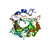
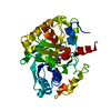
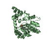
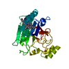
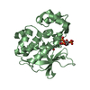
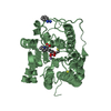
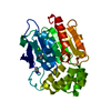
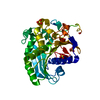
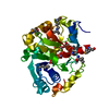
Structural insights into substrate binding of brown spider venom class II phospholipases D
Components
Phospholipase D LiSicTox-alphaIA1bii
Keywords
HYDROLASE / TIM-barrel fold
Function / homology
Function and homology information
phosphoric diester hydrolase activity / lipid catabolic process / Lyases; Phosphorus-oxygen lyases / toxin activity / killing of cells of another organism / lyase activity / extracellular region / metal ion binding Similarity search - Function
Glycerophosphoryl diester phosphodiesterase family / Phosphatidylinositol (PI) phosphodiesterase / PLC-like phosphodiesterase, TIM beta/alpha-barrel domain superfamily / TIM Barrel / Alpha-Beta Barrel / Alpha Beta Similarity search - Domain/homology
Monochromator: Si(111) double-crystal / Protocol: SINGLE WAVELENGTH / Monochromatic (M) / Laue (L): M / Scattering type: x-ray
Radiation wavelength
Wavelength: 1.458 Å / Relative weight: 1
Reflection
Resolution: 1.6→24.49 Å / Num. all: 47089
-
Processing
Software
Name
Version
Classification
HKL-2000
datacollection
MOLREP
phasing
REFMAC
5.8.0049
refinement
DENZO
datareduction
SCALEPACK
datascaling
Refinement
Method to determine structure: MOLECULAR REPLACEMENT / Resolution: 1.64→24.49 Å / Cor.coef. Fo:Fc: 0.963 / Cor.coef. Fo:Fc free: 0.95 / SU B: 2.097 / SU ML: 0.072 / Cross valid method: THROUGHOUT / ESU R: 0.105 / ESU R Free: 0.104 / Stereochemistry target values: MAXIMUM LIKELIHOOD / Details: HYDROGENS HAVE BEEN USED IF PRESENT IN THE INPUT
Rfactor
Num. reflection
% reflection
Selection details
Rfree
0.21592
1569
5.1 %
RANDOM
Rwork
0.17965
-
-
-
obs
0.18156
29302
95.54 %
-
Solvent computation
Ion probe radii: 0.8 Å / Shrinkage radii: 0.8 Å / VDW probe radii: 1.2 Å / Solvent model: MASK
 Movie
Movie Controller

 Yorodumi
 Open data
 Basic information
 Components
 Keywords
 Function and homology information
 Loxosceles intermedia (spider)
 X-RAY DIFFRACTION /
  Authors
 Citation
 Structure visualization
 Downloads & links
 Other downloads
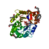

 PDBj
PDBj


 Assembly
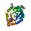


 Mass: 24.305 Da / Num. of mol.: 1 / Source method: obtained synthetically / Formula: Mg
 Mass: 256.424 Da / Num. of mol.: 1 / Source method: obtained synthetically / Formula: C16H32O2
 Mass: 92.094 Da / Num. of mol.: 1 / Source method: obtained synthetically / Formula: C3H8O3
 Mass: 214.344 Da / Num. of mol.: 1 / Source method: obtained synthetically / Formula: C13H26O2
 Mass: 144.211 Da / Num. of mol.: 1 / Source method: obtained synthetically / Formula: C8H16O2
 Sample preparation
 / Beamline: W01B-MX2 / Wavelength: 1.458 Å
 Processing