 Movie
Movie Controller

[English] 日本語
 Yorodumi
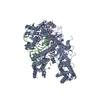
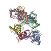
- PDB-4rul: Crystal structure of full-length E.Coli topoisomerase I in comple... -
+ Open data

- Basic information
| Entry | Database: PDB / ID: 4rul | ||||||
|---|---|---|---|---|---|---|---|
| Title | Crystal structure of full-length E.Coli topoisomerase I in complex with ssDNA | ||||||
 Components Components |
| ||||||
 Keywords Keywords | ISOMERASE/DNA / TOPOISOMERASE 1A / ISOMERASE-DNA complex | ||||||
| Function / homology |  Function and homology information Function and homology informationRNA topoisomerase activity / DNA topoisomerase activity / DNA topoisomerase / DNA topoisomerase type I (single strand cut, ATP-independent) activity / DNA topological change / chromosome segregation / chromosome / DNA binding / zinc ion binding / cytosol Similarity search - Function | ||||||
| Biological species |  synthetic construct (others) | ||||||
| Method |  X-RAY DIFFRACTION / SYNCHROTRON / MOLECULAR REPLACEMENT / Resolution: 2.9 Å X-RAY DIFFRACTION / SYNCHROTRON / MOLECULAR REPLACEMENT / Resolution: 2.9 Å | ||||||
 Authors Authors | Tan, K. / Chen, B. / Tse-Dinh, Y.C. | ||||||
 Citation Citation | Journal: Nucleic Acids Res. / Year: 2015 Title: Structural basis for suppression of hypernegative DNA supercoiling by E. coli topoisomerase I. Authors: Tan, K. / Zhou, Q. / Cheng, B. / Zhang, Z. / Joachimiak, A. / Tse-Dinh, Y.C. | ||||||
| History |
|
- Structure visualization
| Structure viewer | Molecule: MolmilJmol/JSmol |
|---|
- Downloads & links
-Download
| PDBx/mmCIF format | 4rul.cif.gz | 348.5 KB | Display | PDBx/mmCIF format |
|---|---|---|---|---|
| PDB format | pdb4rul.ent.gz | 277.6 KB | Display | PDB format |
| PDBx/mmJSON format | 4rul.json.gz | Tree view | PDBx/mmJSON format | |
| Others |  Other downloads Other downloads |
-Validation report
| Arichive directory | https://data.pdbj.org/pub/pdb/validation_reports/ru/4rulftp://data.pdbj.org/pub/pdb/validation_reports/ru/4rul | HTTPS FTP |
|---|
-Related structure data
| Related structure data | 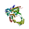 3pwtS S: Starting model for refinement |
|---|---|
| Similar structure data |


-Links
 PDBj
PDBj


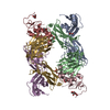
- Assembly
| Deposited unit | 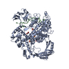
| ||||||||
|---|---|---|---|---|---|---|---|---|---|
| 1 |
| ||||||||
| Unit cell |
|
-Components
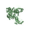
-Protein / DNA chain , 2 types, 2 molecules AB
| #1: Protein | Mass: 97635.047 Da / Num. of mol.: 1 Source method: isolated from a genetically manipulated source Source: (gene. exp.) References: UniProt: C9QXS7, UniProt: P06612*PLUS, EC: 5.99.1.2 |
|---|---|
| #2: DNA chain | Mass: 8997.819 Da / Num. of mol.: 1 / Source method: obtained synthetically / Source: (synth.) synthetic construct (others) |
-Non-polymers , 4 types, 47 molecules 


| #3: Chemical | ChemComp-ZN /  Mass: 65.409 Da / Num. of mol.: 4 / Source method: obtained synthetically / Formula: Zn Mass: 65.409 Da / Num. of mol.: 4 / Source method: obtained synthetically / Formula: Zn#4: Chemical | ChemComp-SO4 /  Mass: 96.063 Da / Num. of mol.: 4 / Source method: obtained synthetically / Formula: SO4 Mass: 96.063 Da / Num. of mol.: 4 / Source method: obtained synthetically / Formula: SO4#5: Chemical | ChemComp-GOL / |  Mass: 92.094 Da / Num. of mol.: 1 / Source method: obtained synthetically / Formula: C3H8O3 Mass: 92.094 Da / Num. of mol.: 1 / Source method: obtained synthetically / Formula: C3H8O3#6: Water | ChemComp-HOH / | Mass: 18.015 Da / Num. of mol.: 38 / Source method: isolated from a natural source / Formula: H2O |
|---|
-Experimental details
-Experiment
| Experiment | Method: X-RAY DIFFRACTION / Number of used crystals: 1 |
|---|
- Sample preparation
| Crystal | Density Matthews: 3.15 Å3/Da / Density % sol: 60.89 % |
|---|---|
| Crystal grow | Temperature: 297 K / Method: vapor diffusion, sitting drop / pH: 6 Details: 0.125M ammonium sulfate, 0.1M MES, 1mM zinc sulfate, 19% PEG 5000 monomethyl ether, pH 6.0, VAPOR DIFFUSION, SITTING DROP, temperature 297K |
-Data collection
| Diffraction | Mean temperature: 100 K |
|---|---|
| Diffraction source | Source: SYNCHROTRON / Site: APS  / Beamline: 19-ID / Wavelength: 0.97913 Å / Beamline: 19-ID / Wavelength: 0.97913 Å |
| Detector | Type: ADSC QUANTUM 315r / Detector: CCD / Date: Mar 17, 2014 / Details: mirror |
| Radiation | Monochromator: Si 111 crystal / Protocol: SINGLE WAVELENGTH / Monochromatic (M) / Laue (L): M / Scattering type: x-ray |
| Radiation wavelength | Wavelength: 0.97913 Å / Relative weight: 1 |
| Reflection | Resolution: 2.9→38 Å / Num. all: 29533 / Num. obs: 29533 / % possible obs: 99.6 % / Observed criterion σ(F): 0 / Observed criterion σ(I): -5 / Redundancy: 3.5 % / Biso Wilson estimate: 66.4 Å2 / Rmerge(I) obs: 0.064 / Net I/σ(I): 23.4 |
| Reflection shell | Resolution: 2.9→2.95 Å / Redundancy: 3.6 % / Rmerge(I) obs: 0.676 / Mean I/σ(I) obs: 1.9 / % possible all: 100 |
- Processing
| Software |
| |||||||||||||||||||||||||||||||||||||||||||||||||||||||||||||||||||||||||||||||||||||||||||||||||||||||||||||||||||||||||||||
|---|---|---|---|---|---|---|---|---|---|---|---|---|---|---|---|---|---|---|---|---|---|---|---|---|---|---|---|---|---|---|---|---|---|---|---|---|---|---|---|---|---|---|---|---|---|---|---|---|---|---|---|---|---|---|---|---|---|---|---|---|---|---|---|---|---|---|---|---|---|---|---|---|---|---|---|---|---|---|---|---|---|---|---|---|---|---|---|---|---|---|---|---|---|---|---|---|---|---|---|---|---|---|---|---|---|---|---|---|---|---|---|---|---|---|---|---|---|---|---|---|---|---|---|---|---|---|
| Refinement | Method to determine structure: MOLECULAR REPLACEMENT Starting model: PDB ENTRY: 3PWT Resolution: 2.9→37.735 Å / SU ML: 0.36 / σ(F): 1.35 / Phase error: 26.79 / Stereochemistry target values: ML
| |||||||||||||||||||||||||||||||||||||||||||||||||||||||||||||||||||||||||||||||||||||||||||||||||||||||||||||||||||||||||||||
| Solvent computation | Shrinkage radii: 0.9 Å / VDW probe radii: 1.11 Å / Solvent model: FLAT BULK SOLVENT MODEL | |||||||||||||||||||||||||||||||||||||||||||||||||||||||||||||||||||||||||||||||||||||||||||||||||||||||||||||||||||||||||||||
| Refinement step | Cycle: LAST / Resolution: 2.9→37.735 Å
| |||||||||||||||||||||||||||||||||||||||||||||||||||||||||||||||||||||||||||||||||||||||||||||||||||||||||||||||||||||||||||||
| Refine LS restraints |
| |||||||||||||||||||||||||||||||||||||||||||||||||||||||||||||||||||||||||||||||||||||||||||||||||||||||||||||||||||||||||||||
| LS refinement shell |
| |||||||||||||||||||||||||||||||||||||||||||||||||||||||||||||||||||||||||||||||||||||||||||||||||||||||||||||||||||||||||||||
| Refinement TLS params. | Method: refined / Refine-ID: X-RAY DIFFRACTION
| |||||||||||||||||||||||||||||||||||||||||||||||||||||||||||||||||||||||||||||||||||||||||||||||||||||||||||||||||||||||||||||
| Refinement TLS group |
|
