 Movie
Movie Controller

[English] 日本語
 Yorodumi
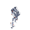
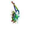
- PDB-4ru3: Crystal structure of the cell puncturing protein Orf41 from Pseud... -
+ Open data

- Basic information
| Entry | Database: PDB / ID: 4ru3 | ||||||
|---|---|---|---|---|---|---|---|
| Title | Crystal structure of the cell puncturing protein Orf41 from Pseudomonas phage SN | ||||||
 Components Components | puncturing protein gp41 | ||||||
 Keywords Keywords | CELL INVASION / gp41 / membrane translocation / phage baseplate / Cell puncturing device | ||||||
| Function / homology | Phage protein Gp138 N-terminal domain / Phage protein Gp138 N-terminal domain / Vgr protein, OB-fold domain superfamily / metal ion binding / : / Putative baseplate protein Function and homology information Function and homology information | ||||||
| Biological species |  Pseudomonas phage SN (virus) Pseudomonas phage SN (virus) | ||||||
| Method |  X-RAY DIFFRACTION / SYNCHROTRON / MOLECULAR REPLACEMENT / Resolution: 1.172 Å X-RAY DIFFRACTION / SYNCHROTRON / MOLECULAR REPLACEMENT / Resolution: 1.172 Å | ||||||
 Authors Authors | Browning, C. / Shneider, M.M. / Leiman, P.G. | ||||||
 Citation Citation | Journal: To be Published Title: Crystal structure of the cell puncturing protein Orf41 from Pseudomonas phage SN Authors: Browning, C. / Shneider, M.M. / Leiman, P.G. | ||||||
| History |
|
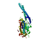
- Structure visualization
| Structure viewer | Molecule: MolmilJmol/JSmol |
|---|
- Downloads & links
-Download
| PDBx/mmCIF format | 4ru3.cif.gz | 113.8 KB | Display | PDBx/mmCIF format |
|---|---|---|---|---|
| PDB format | pdb4ru3.ent.gz | 88.5 KB | Display | PDB format |
| PDBx/mmJSON format | 4ru3.json.gz | Tree view | PDBx/mmJSON format | |
| Others |  Other downloads Other downloads |
-Validation report
| Summary document | 4ru3_validation.pdf.gz | 442.4 KB | Display | wwPDB validaton report |
|---|---|---|---|---|
| Full document | 4ru3_full_validation.pdf.gz | 446.8 KB | Display | |
| Data in XML | 4ru3_validation.xml.gz | 13.7 KB | Display | |
| Data in CIF | 4ru3_validation.cif.gz | 20.3 KB | Display | |
| Arichive directory | https://data.pdbj.org/pub/pdb/validation_reports/ru/4ru3ftp://data.pdbj.org/pub/pdb/validation_reports/ru/4ru3 | HTTPS FTP |
-Related structure data
| Similar structure data |
|---|


-Links
 PDBj
PDBj- Assembly
| Deposited unit | 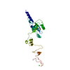
| |||||||||
|---|---|---|---|---|---|---|---|---|---|---|
| 1 | 
| |||||||||
| Unit cell |
| |||||||||
| Components on special symmetry positions |
|
-Components
| #1: Protein | Mass: 24085.992 Da / Num. of mol.: 1 Source method: isolated from a genetically manipulated source Source: (gene. exp.) Pseudomonas phage SN (virus) / Gene: ORF41Plasmid: pEE3, pET-23 derivative (TEV cleavage site after His-tag) Production host:  |
|---|---|
| #2: Chemical | ChemComp-FE /   Mass: 55.845 Da / Num. of mol.: 1 / Source method: obtained synthetically / Formula: Fe Mass: 55.845 Da / Num. of mol.: 1 / Source method: obtained synthetically / Formula: Fe |
| #3: Chemical | ChemComp-CA /   Mass: 40.078 Da / Num. of mol.: 1 / Source method: obtained synthetically / Formula: Ca Mass: 40.078 Da / Num. of mol.: 1 / Source method: obtained synthetically / Formula: Ca |
| #4: Chemical | ChemComp-GOL /   Mass: 92.094 Da / Num. of mol.: 1 / Source method: obtained synthetically / Formula: C3H8O3 Mass: 92.094 Da / Num. of mol.: 1 / Source method: obtained synthetically / Formula: C3H8O3 |
| #5: Water | ChemComp-HOH /  Mass: 18.015 Da / Num. of mol.: 310 / Source method: isolated from a natural source / Formula: H2O Mass: 18.015 Da / Num. of mol.: 310 / Source method: isolated from a natural source / Formula: H2O |
-Experimental details
-Experiment
| Experiment | Method: X-RAY DIFFRACTION / Number of used crystals: 1 |
|---|
- Sample preparation
| Crystal | Density Matthews: 2.09 Å3/Da / Density % sol: 41.16 % |
|---|---|
| Crystal grow | Temperature: 291 K / Method: vapor diffusion / pH: 5 Details: 21% PEG 10K, 0.1M Ammonium acetate, 0.1M Bis-Tris pH 5.0, VAPOR DIFFUSION, temperature 291K |
-Data collection
| Diffraction | Mean temperature: 100 K |
|---|---|
| Diffraction source | Source: SYNCHROTRON / Site: SLS  / Beamline: X06SA / Wavelength: 1 Å / Beamline: X06SA / Wavelength: 1 Å |
| Detector | Type: DECTRIS PILATUS 6M / Detector: PIXEL / Date: Sep 5, 2011 |
| Radiation | Monochromator: Si(111) Monochromator / Protocol: SINGLE WAVELENGTH / Monochromatic (M) / Laue (L): M / Scattering type: x-ray |
| Radiation wavelength | Wavelength: 1 Å / Relative weight: 1 |
| Reflection | Resolution: 1.17→34.513 Å / Num. all: 67585 / Num. obs: 67579 / % possible obs: 100 % / Observed criterion σ(F): 0 / Observed criterion σ(I): 2 / Redundancy: 17.7 % / Rmerge(I) obs: 0.068 / Rsym value: 0.071 / Net I/σ(I): 23.1 |
| Reflection shell | Resolution: 1.17→1.24 Å / Redundancy: 17.8 % / Rmerge(I) obs: 0.293 / Mean I/σ(I) obs: 8.8 / Num. unique all: 9781 / Rsym value: 0.309 / % possible all: 100 |
- Processing
| Software |
| |||||||||||||||||||||||||||||||||||||||||||||||||||||||||||||||||||||||||||||||||||||||||||||||||||||||||
|---|---|---|---|---|---|---|---|---|---|---|---|---|---|---|---|---|---|---|---|---|---|---|---|---|---|---|---|---|---|---|---|---|---|---|---|---|---|---|---|---|---|---|---|---|---|---|---|---|---|---|---|---|---|---|---|---|---|---|---|---|---|---|---|---|---|---|---|---|---|---|---|---|---|---|---|---|---|---|---|---|---|---|---|---|---|---|---|---|---|---|---|---|---|---|---|---|---|---|---|---|---|---|---|---|---|---|
| Refinement | Method to determine structure: MOLECULAR REPLACEMENT / Resolution: 1.172→34.513 Å / SU ML: 0.07 / σ(F): 0.08 / Phase error: 11.31 / Stereochemistry target values: Engh & Huber
| |||||||||||||||||||||||||||||||||||||||||||||||||||||||||||||||||||||||||||||||||||||||||||||||||||||||||
| Solvent computation | Shrinkage radii: 0.9 Å / VDW probe radii: 1.11 Å / Solvent model: FLAT BULK SOLVENT MODEL | |||||||||||||||||||||||||||||||||||||||||||||||||||||||||||||||||||||||||||||||||||||||||||||||||||||||||
| Refinement step | Cycle: LAST / Resolution: 1.172→34.513 Å
| |||||||||||||||||||||||||||||||||||||||||||||||||||||||||||||||||||||||||||||||||||||||||||||||||||||||||
| Refine LS restraints |
| |||||||||||||||||||||||||||||||||||||||||||||||||||||||||||||||||||||||||||||||||||||||||||||||||||||||||
| LS refinement shell |
|
