 Movie
Movie Controller

[English] 日本語
 Yorodumi
- PDB-4rnd: Crystal Structure of the subunit DF-assembly of the eukaryotic V-... -
+ Open data

- Basic information
| Entry | Database: PDB / ID: 4rnd | ||||||
|---|---|---|---|---|---|---|---|
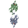
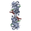
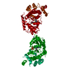
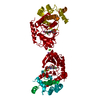
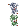
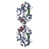
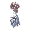
| Title | Crystal Structure of the subunit DF-assembly of the eukaryotic V-ATPase. | ||||||
 Components Components |
| ||||||
 Keywords Keywords | HYDROLASE / alpha helical / Rossmann Fold / Regulatory / Coupling | ||||||
| Function / homology |  Function and homology information Function and homology informationInsulin receptor recycling / Transferrin endocytosis and recycling / ROS and RNS production in phagocytes / Amino acids regulate mTORC1 / Golgi lumen acidification / vacuolar proton-transporting V-type ATPase, V1 domain / endosomal lumen acidification / proton-transporting V-type ATPase complex / vacuolar proton-transporting V-type ATPase complex / vacuolar acidification ...Insulin receptor recycling / Transferrin endocytosis and recycling / ROS and RNS production in phagocytes / Amino acids regulate mTORC1 / Golgi lumen acidification / vacuolar proton-transporting V-type ATPase, V1 domain / endosomal lumen acidification / proton-transporting V-type ATPase complex / vacuolar proton-transporting V-type ATPase complex / vacuolar acidification / fungal-type vacuole membrane / proton-transporting ATPase activity, rotational mechanism / Neutrophil degranulation / proton transmembrane transport / membrane raft / Golgi membrane / membrane Similarity search - Function | ||||||
| Biological species |  | ||||||
| Method |  X-RAY DIFFRACTION / SYNCHROTRON / SAD / Resolution: 3.18 Å X-RAY DIFFRACTION / SYNCHROTRON / SAD / Resolution: 3.18 Å | ||||||
 Authors Authors | Balakrishna, A.M. / Basak, S. / Gruber, G. | ||||||
 Citation Citation | Journal: J.Biol.Chem. / Year: 2015 Title: Crystal Structure of Subunits D and F in Complex Gives Insight into Energy Transmission of the Eukaryotic V-ATPase from Saccharomyces cerevisiae. Authors: Balakrishna, A.M. / Basak, S. / Manimekalai, M.S. / Gruber, G. #1: Journal: J.Biol.Chem. / Year: 2013Title: Crystal and NMR structures give insights into the role and dynamics of subunit F of the eukaryotic V-ATPase from Saccharomyces cerevisiae. Authors: Basak, S. / Lim, J. / Manimekalai, M.S. / Balakrishna, A.M. / Gruber, G. | ||||||
| History |
|
- Structure visualization
| Structure viewer | Molecule: MolmilJmol/JSmol |
|---|
- Downloads & links
-Download
| PDBx/mmCIF format | 4rnd.cif.gz | 241.9 KB | Display | PDBx/mmCIF format |
|---|---|---|---|---|
| PDB format | pdb4rnd.ent.gz | 199.1 KB | Display | PDB format |
| PDBx/mmJSON format | 4rnd.json.gz | Tree view | PDBx/mmJSON format | |
| Others |  Other downloads Other downloads |
-Validation report
| Summary document | 4rnd_validation.pdf.gz | 460.5 KB | Display | wwPDB validaton report |
|---|---|---|---|---|
| Full document | 4rnd_full_validation.pdf.gz | 463.2 KB | Display | |
| Data in XML | 4rnd_validation.xml.gz | 22.1 KB | Display | |
| Data in CIF | 4rnd_validation.cif.gz | 30.1 KB | Display | |
| Arichive directory | https://data.pdbj.org/pub/pdb/validation_reports/rn/4rndftp://data.pdbj.org/pub/pdb/validation_reports/rn/4rnd | HTTPS FTP |
-Related structure data
| Related structure data | |
|---|---|
| Similar structure data |


-Links
 PDBj
PDBj


- Assembly
| Deposited unit | 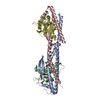
| ||||||||||||||||||||||||||||||||||||||||||||||||||||||||||||||||||||
|---|---|---|---|---|---|---|---|---|---|---|---|---|---|---|---|---|---|---|---|---|---|---|---|---|---|---|---|---|---|---|---|---|---|---|---|---|---|---|---|---|---|---|---|---|---|---|---|---|---|---|---|---|---|---|---|---|---|---|---|---|---|---|---|---|---|---|---|---|---|
| 1 |
| ||||||||||||||||||||||||||||||||||||||||||||||||||||||||||||||||||||
| Unit cell |
| ||||||||||||||||||||||||||||||||||||||||||||||||||||||||||||||||||||
| Noncrystallographic symmetry (NCS) | NCS domain:
NCS domain segments: Component-ID: _ / Refine code: _
NCS ensembles :
|
-Components
| #1: Protein | Mass: 29235.023 Da / Num. of mol.: 2 Source method: isolated from a genetically manipulated source Source: (gene. exp.)  #2: Protein | Mass: 13479.170 Da / Num. of mol.: 2 Source method: isolated from a genetically manipulated source Source: (gene. exp.) #3: Chemical |   Mass: 92.094 Da / Num. of mol.: 3 / Source method: obtained synthetically / Formula: C3H8O3 Mass: 92.094 Da / Num. of mol.: 3 / Source method: obtained synthetically / Formula: C3H8O3#4: Water | ChemComp-HOH / |  Mass: 18.015 Da / Num. of mol.: 59 / Source method: isolated from a natural source / Formula: H2O Mass: 18.015 Da / Num. of mol.: 59 / Source method: isolated from a natural source / Formula: H2O |
|---|
-Experimental details
-Experiment
| Experiment | Method: X-RAY DIFFRACTION / Number of used crystals: 1 |
|---|
- Sample preparation
| Crystal | Density Matthews: 6.14 Å3/Da / Density % sol: 79.96 % |
|---|---|
| Crystal grow | Temperature: 291 K / Method: vapor diffusion, hanging drop / pH: 5.6 Details: 0.1 M sodium citrate tribasic dehydrate, 1.2 M Ammonium citrate monobasic, ph 5.6, VAPOR DIFFUSION, HANGING DROP, temperature 291K |
-Data collection
| Diffraction | Mean temperature: 100 K |
|---|---|
| Diffraction source | Source: SYNCHROTRON / Site: NSRRC  / Beamline: BL13B1 / Wavelength: 0.978 Å / Beamline: BL13B1 / Wavelength: 0.978 Å |
| Detector | Type: ADSC QUANTUM 315 / Detector: CCD / Date: May 22, 2014 Details: Vertically Collimating Premirror, Toroidal Focusing Mirror |
| Radiation | Monochromator: Double Crystal Si(111) Monochromator / Protocol: SINGLE WAVELENGTH / Monochromatic (M) / Laue (L): M / Scattering type: x-ray |
| Radiation wavelength | Wavelength: 0.978 Å / Relative weight: 1 |
| Reflection | Resolution: 3.18→30 Å / Num. all: 38345 / Num. obs: 33061 / % possible obs: 80.08 % / Observed criterion σ(F): 0 / Observed criterion σ(I): 2 / Redundancy: 11.8 % / Rmerge(I) obs: 0.09 / Net I/σ(I): 19.1 |
| Reflection shell | Resolution: 3.1→3.21 Å / Redundancy: 8.7 % / Rmerge(I) obs: 0.84 / Mean I/σ(I) obs: 1.2 / % possible all: 98.3 |
- Processing
| Software |
| ||||||||||||||||||||||||||||||||||||||||||||||||||||||||||||||||||||||||||||||||||||||||||||||||||||||||||||||||||||||||||||||||||||||||||||||||||||||||||||||||||||||||||||||||||||||
|---|---|---|---|---|---|---|---|---|---|---|---|---|---|---|---|---|---|---|---|---|---|---|---|---|---|---|---|---|---|---|---|---|---|---|---|---|---|---|---|---|---|---|---|---|---|---|---|---|---|---|---|---|---|---|---|---|---|---|---|---|---|---|---|---|---|---|---|---|---|---|---|---|---|---|---|---|---|---|---|---|---|---|---|---|---|---|---|---|---|---|---|---|---|---|---|---|---|---|---|---|---|---|---|---|---|---|---|---|---|---|---|---|---|---|---|---|---|---|---|---|---|---|---|---|---|---|---|---|---|---|---|---|---|---|---|---|---|---|---|---|---|---|---|---|---|---|---|---|---|---|---|---|---|---|---|---|---|---|---|---|---|---|---|---|---|---|---|---|---|---|---|---|---|---|---|---|---|---|---|---|---|---|---|
| Refinement | Method to determine structure: SAD / Resolution: 3.18→29.63 Å / Cor.coef. Fo:Fc: 0.92 / Cor.coef. Fo:Fc free: 0.895 / SU B: 20.993 / SU ML: 0.174 / Cross valid method: THROUGHOUT / ESU R: 0.082 / ESU R Free: 0.061 / Stereochemistry target values: MAXIMUM LIKELIHOOD / Details: HYDROGENS HAVE BEEN ADDED IN THE RIDING POSITIONS
| ||||||||||||||||||||||||||||||||||||||||||||||||||||||||||||||||||||||||||||||||||||||||||||||||||||||||||||||||||||||||||||||||||||||||||||||||||||||||||||||||||||||||||||||||||||||
| Solvent computation | Ion probe radii: 0.8 Å / Shrinkage radii: 0.8 Å / VDW probe radii: 1.2 Å / Solvent model: MASK | ||||||||||||||||||||||||||||||||||||||||||||||||||||||||||||||||||||||||||||||||||||||||||||||||||||||||||||||||||||||||||||||||||||||||||||||||||||||||||||||||||||||||||||||||||||||
| Displacement parameters | Biso mean: 52.757 Å2
| ||||||||||||||||||||||||||||||||||||||||||||||||||||||||||||||||||||||||||||||||||||||||||||||||||||||||||||||||||||||||||||||||||||||||||||||||||||||||||||||||||||||||||||||||||||||
| Refinement step | Cycle: LAST / Resolution: 3.18→29.63 Å
| ||||||||||||||||||||||||||||||||||||||||||||||||||||||||||||||||||||||||||||||||||||||||||||||||||||||||||||||||||||||||||||||||||||||||||||||||||||||||||||||||||||||||||||||||||||||
| Refine LS restraints |
|