 Movie
Movie Controller

[English] 日本語
 Yorodumi
- PDB-1dp5: THE STRUCTURE OF PROTEINASE A COMPLEXED WITH A IA3 MUTANT INHIBITOR -
+ Open data

- Basic information
| Entry | Database: PDB / ID: 1dp5 | |||||||||
|---|---|---|---|---|---|---|---|---|---|---|
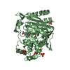
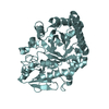
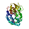
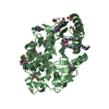
| Title | THE STRUCTURE OF PROTEINASE A COMPLEXED WITH A IA3 MUTANT INHIBITOR | |||||||||
 Components Components |
| |||||||||
 Keywords Keywords | HYDROLASE/HYDROLASE INHIBITOR / Proteinase A / MMM / IA3 mutant / HYDROLASE-HYDROLASE INHIBITOR COMPLEX | |||||||||
| Function / homology |  Function and homology information Function and homology informationsaccharopepsin / protein catabolic process in the vacuole / microautophagy / cytoplasm to vacuole targeting by the Cvt pathway / oligosaccharide binding / aspartic-type endopeptidase inhibitor activity / fungal-type vacuole / pexophagy / vacuole / endopeptidase inhibitor activity ...saccharopepsin / protein catabolic process in the vacuole / microautophagy / cytoplasm to vacuole targeting by the Cvt pathway / oligosaccharide binding / aspartic-type endopeptidase inhibitor activity / fungal-type vacuole / pexophagy / vacuole / endopeptidase inhibitor activity / : / macroautophagy / autophagy / disordered domain specific binding / peptidase activity / protease binding / aspartic-type endopeptidase activity / endoplasmic reticulum / protein-containing complex / mitochondrion / nucleus / cytoplasm Similarity search - Function | |||||||||
| Biological species |  | |||||||||
| Method |  X-RAY DIFFRACTION / SYNCHROTRON / Resolution: 2.2 Å X-RAY DIFFRACTION / SYNCHROTRON / Resolution: 2.2 Å | |||||||||
 Authors Authors | Li, M. / Phylip, H.L. / Lees, W.E. / Winther, J.R. / Dunn, B.M. / Wlodawer, A. / Kay, J. / Guschina, A. | |||||||||
 Citation Citation | Journal: Nat.Struct.Biol. / Year: 2000 Title: The aspartic proteinase from Saccharomyces cerevisiae folds its own inhibitor into a helix. Authors: Li, M. / Phylip, L.H. / Lees, W.E. / Winther, J.R. / Dunn, B.M. / Wlodawer, A. / Kay, J. / Gustchina, A. #1: Journal: J.Mol.Biol. / Year: 1997Title: The Three-dimensional Structure at 2.4 A Resolution of Glycosylated Proteinase A from the Lysosome-like Vacuole of Saccharomyces Cerevisiae. Authors: Aguilar, C.F. / Cronin, N.B. / Badasso, M. / Dreyer, T. / Newman, M.P. / Cooper, J.B. / Hoover, D.J. / Wood, S.P. / Johnson, M.S. / Blundell, T.L. | |||||||||
| History |
|
- Structure visualization
| Structure viewer | Molecule: MolmilJmol/JSmol |
|---|
- Downloads & links
-Download
| PDBx/mmCIF format | 1dp5.cif.gz | 96.6 KB | Display | PDBx/mmCIF format |
|---|---|---|---|---|
| PDB format | pdb1dp5.ent.gz | 71.8 KB | Display | PDB format |
| PDBx/mmJSON format | 1dp5.json.gz | Tree view | PDBx/mmJSON format | |
| Others |  Other downloads Other downloads |
-Validation report
| Arichive directory | https://data.pdbj.org/pub/pdb/validation_reports/dp/1dp5ftp://data.pdbj.org/pub/pdb/validation_reports/dp/1dp5 | HTTPS FTP |
|---|
-Related structure data


-Links
 PDBj
PDBj- Assembly
| Deposited unit | 
| ||||||||
|---|---|---|---|---|---|---|---|---|---|
| 1 |
| ||||||||
| 2 | 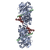
| ||||||||
| 3 | 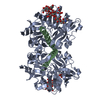
| ||||||||
| Unit cell |
|
-Components
| #1: Protein | Mass: 35774.551 Da / Num. of mol.: 1 / Source method: isolated from a natural source / Source: (natural) |
|---|---|
| #2: Protein | Mass: 7726.512 Da / Num. of mol.: 1 / Mutation: K31M, K32M Source method: isolated from a genetically manipulated source Source: (gene. exp.) Production host:  |
| #3: Polysaccharide | beta-D-mannopyranose-(1-2)-alpha-D-mannopyranose-(1-2)-[alpha-D-mannopyranose-(1-6)]alpha-D- ...beta-D-mannopyranose-(1-2)-alpha-D-mannopyranose-(1-2)-[alpha-D-mannopyranose-(1-6)]alpha-D-mannopyranose-(1-3)-[beta-D-mannopyranose-(1-6)-alpha-D-mannopyranose-(1-6)]beta-D-mannopyranose-(1-4)-2-acetamido-2-deoxy-beta-D-glucopyranose-(1-4)-2-acetamido-2-deoxy-beta-D-glucopyranose Source method: isolated from a genetically manipulated source |
| #4: Water | ChemComp-HOH /  Mass: 18.015 Da / Num. of mol.: 391 / Source method: isolated from a natural source / Formula: H2O Mass: 18.015 Da / Num. of mol.: 391 / Source method: isolated from a natural source / Formula: H2O |
| Has protein modification | Y |
| Sequence details | THE RESIDUE NUMBERS FOR CHAIN A FOLLOW THE RESIDUE NUMBERS IN PDB ENTRY 2JXR. THE SEQUENCE FOR ...THE RESIDUE NUMBERS FOR CHAIN A FOLLOW THE RESIDUE NUMBERS IN PDB ENTRY 2JXR. THE SEQUENCE FOR CHAIN B IS THE FULL LENGTH SEQUENCE. |
-Experimental details
-Experiment
| Experiment | Method: X-RAY DIFFRACTION / Number of used crystals: 1 |
|---|
- Sample preparation
| Crystal | Density Matthews: 3.21 Å3/Da / Density % sol: 61.63 % | ||||||||||||||||||||||||||||||
|---|---|---|---|---|---|---|---|---|---|---|---|---|---|---|---|---|---|---|---|---|---|---|---|---|---|---|---|---|---|---|---|
| Crystal grow | Temperature: 298 K / Method: vapor diffusion, hanging drop / pH: 5.6 Details: PEG 1500, Ammounia Sulfate, pH 5.6, VAPOR DIFFUSION, HANGING DROP, temperature 298.K | ||||||||||||||||||||||||||||||
| Crystal grow | *PLUS pH: 6.6 | ||||||||||||||||||||||||||||||
| Components of the solutions | *PLUS
|
-Data collection
| Diffraction | Mean temperature: 298 K |
|---|---|
| Diffraction source | Source: SYNCHROTRON / Site: NSLS  / Beamline: X9B / Wavelength: 1.54 / Beamline: X9B / Wavelength: 1.54 |
| Detector | Type: ADSC QUANTUM / Detector: CCD / Date: Jan 1, 1999 |
| Radiation | Protocol: SINGLE WAVELENGTH / Monochromatic (M) / Laue (L): M / Scattering type: x-ray |
| Radiation wavelength | Wavelength: 1.54 Å / Relative weight: 1 |
| Reflection | Resolution: 2.2→20 Å / Num. all: 29595 / Num. obs: 28115 / % possible obs: 95 % / Observed criterion σ(F): 0 / Observed criterion σ(I): 0 / Redundancy: 5.6 % / Rmerge(I) obs: 0.069 / Net I/σ(I): 16 |
| Reflection shell | Resolution: 2.2→2.28 Å / Redundancy: 5.7 % / Rmerge(I) obs: 0.332 / Num. unique all: 2881 / % possible all: 99.6 |
| Reflection | *PLUS % possible obs: 95.6 % / Redundancy: 5.8 % |
| Reflection shell | *PLUS % possible obs: 99.6 % / Mean I/σ(I) obs: 6 |
- Processing
| Software |
| |||||||||||||||||||||||||
|---|---|---|---|---|---|---|---|---|---|---|---|---|---|---|---|---|---|---|---|---|---|---|---|---|---|---|
| Refinement | Resolution: 2.2→30 Å / σ(F): 0 / σ(I): 0 / Stereochemistry target values: Engh & Huber / Details: Residues 33-68 are disordered.
| |||||||||||||||||||||||||
| Refinement step | Cycle: LAST / Resolution: 2.2→30 Å
| |||||||||||||||||||||||||
| Refine LS restraints |
| |||||||||||||||||||||||||
| Software | *PLUS Name: CNS / Classification: refinement | |||||||||||||||||||||||||
| Refinement | *PLUS Highest resolution: 2.2 Å / Lowest resolution: 30 Å / σ(F): 0 / Rfactor obs: 0.1883 | |||||||||||||||||||||||||
| Solvent computation | *PLUS | |||||||||||||||||||||||||
| Displacement parameters | *PLUS |
