 Movie
Movie Controller

[English] 日本語
 Yorodumi
- PDB-4rmb: Crystal structure of keratin 4 binding domain of surface adhesin ... -
+ Open data

- Basic information
| Entry | Database: PDB / ID: 4rmb | ||||||
|---|---|---|---|---|---|---|---|
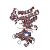
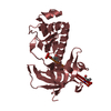
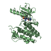
| Title | Crystal structure of keratin 4 binding domain of surface adhesin Srr-1 of S.agalactiae | ||||||
 Components Components | Serine rich repeat protein-1 (Srr-1) | ||||||
 Keywords Keywords | CELL ADHESION / Variant of DEv-IgG fold / adhesin / keratin 4 / bacterial cell surface / beta sheet complementation | ||||||
| Function / homology |  Function and homology information Function and homology informationkeratin filament binding / symbiont entry into host / cell adhesion / cell surface / extracellular region / metal ion binding Similarity search - Function | ||||||
| Biological species |  Streptococcus agalactiae NEM316 (bacteria) Streptococcus agalactiae NEM316 (bacteria) | ||||||
| Method |  X-RAY DIFFRACTION / SYNCHROTRON / MOLECULAR REPLACEMENT / Resolution: 1.7 Å X-RAY DIFFRACTION / SYNCHROTRON / MOLECULAR REPLACEMENT / Resolution: 1.7 Å | ||||||
 Authors Authors | Ponnuraj, K. / Sundaresan, R. | ||||||
 Citation Citation | Journal: Int.J.Biol.Macromol. / Year: 2015 Title: Structure of KRT4 binding domain of Srr-1 from Streptococcus agalactiae reveals a novel beta-sheet complementation. Authors: Sundaresan, R. / Samen, U. / Ponnuraj, K. | ||||||
| History |
|
- Structure visualization
| Structure viewer | Molecule: MolmilJmol/JSmol |
|---|
- Downloads & links
-Download
| PDBx/mmCIF format | 4rmb.cif.gz | 70 KB | Display | PDBx/mmCIF format |
|---|---|---|---|---|
| PDB format | pdb4rmb.ent.gz | 52.4 KB | Display | PDB format |
| PDBx/mmJSON format | 4rmb.json.gz | Tree view | PDBx/mmJSON format | |
| Others |  Other downloads Other downloads |
-Validation report
| Summary document | 4rmb_validation.pdf.gz | 439.6 KB | Display | wwPDB validaton report |
|---|---|---|---|---|
| Full document | 4rmb_full_validation.pdf.gz | 442.7 KB | Display | |
| Data in XML | 4rmb_validation.xml.gz | 13.9 KB | Display | |
| Data in CIF | 4rmb_validation.cif.gz | 18.6 KB | Display | |
| Arichive directory | https://data.pdbj.org/pub/pdb/validation_reports/rm/4rmbftp://data.pdbj.org/pub/pdb/validation_reports/rm/4rmb | HTTPS FTP |
-Related structure data
| Related structure data | 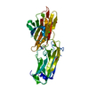 4mboS S: Starting model for refinement |
|---|---|
| Similar structure data |


-Links
 PDBj
PDBj
- Assembly
| Deposited unit | 
| ||||||||
|---|---|---|---|---|---|---|---|---|---|
| 1 |
| ||||||||
| Unit cell |
|
-Components
| #1: Protein | Mass: 18526.262 Da / Num. of mol.: 2 / Fragment: Keratin 4 binding domain Source method: isolated from a genetically manipulated source Source: (gene. exp.) Streptococcus agalactiae NEM316 (bacteria)Strain: NEM316 / Gene: gbs1529 / Plasmid: pET28a / Production host: #2: Water | ChemComp-HOH / |  Mass: 18.015 Da / Num. of mol.: 107 / Source method: isolated from a natural source / Formula: H2O Mass: 18.015 Da / Num. of mol.: 107 / Source method: isolated from a natural source / Formula: H2O |
|---|
-Experimental details
-Experiment
| Experiment | Method: X-RAY DIFFRACTION / Number of used crystals: 1 |
|---|
- Sample preparation
| Crystal | Density Matthews: 1.78 Å3/Da / Density % sol: 31 % |
|---|---|
| Crystal grow | Temperature: 293.15 K / Method: vapor diffusion, hanging drop / pH: 6.7 Details: 25% (w/v) PEG 3350 (Sigma), 100 mM Lithium chloride and 100 mM Imidazole, pH 6.7, VAPOR DIFFUSION, HANGING DROP, temperature 293.15K |
-Data collection
| Diffraction | Mean temperature: 100 K |
|---|---|
| Diffraction source | Source: SYNCHROTRON / Site: ELETTRA  / Beamline: 5.2R / Wavelength: 0.97927 Å / Beamline: 5.2R / Wavelength: 0.97927 Å |
| Detector | Type: DECTRIS PILATUS 2M / Detector: PIXEL / Date: Oct 27, 2013 |
| Radiation | Protocol: SINGLE WAVELENGTH / Monochromatic (M) / Laue (L): M / Scattering type: x-ray |
| Radiation wavelength | Wavelength: 0.97927 Å / Relative weight: 1 |
| Reflection | Resolution: 1.7→35.95 Å / Num. all: 27350 / Num. obs: 27350 / % possible obs: 95.4 % / Observed criterion σ(F): 0 / Observed criterion σ(I): 0 / Redundancy: 5.8 % / Rmerge(I) obs: 0.066 / Net I/σ(I): 14.1 |
| Reflection shell | Resolution: 1.7→1.79 Å / Redundancy: 5.8 % / Rmerge(I) obs: 0.66 / Num. unique all: 27350 / % possible all: 80.7 |
- Processing
| Software |
| |||||||||||||||||||||||||
|---|---|---|---|---|---|---|---|---|---|---|---|---|---|---|---|---|---|---|---|---|---|---|---|---|---|---|
| Refinement | Method to determine structure: MOLECULAR REPLACEMENT Starting model: PDB ENTRY 4MBO Resolution: 1.7→20 Å / Cross valid method: THROUGHOUT / σ(F): 0 / σ(I): 0 / Stereochemistry target values: Engh & Huber
| |||||||||||||||||||||||||
| Displacement parameters | Biso mean: 24.22 Å2
| |||||||||||||||||||||||||
| Refinement step | Cycle: LAST / Resolution: 1.7→20 Å
|
