 Movie
Movie Controller

[English] 日本語
 Yorodumi
- PDB-4rm4: The crystal structure of the versatile cytochrome P450 enzyme CYP... -
+ Open data

- Basic information
| Entry | Database: PDB / ID: 4rm4 | ||||||
|---|---|---|---|---|---|---|---|
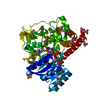
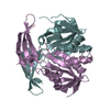
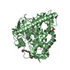
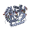
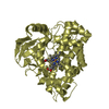
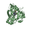
| Title | The crystal structure of the versatile cytochrome P450 enzyme CYP109B1 from Bacillus subtilis | ||||||
 Components Components | Cytochrome P450 | ||||||
 Keywords Keywords | ELECTRON TRANSPORT / Cytochrome P450 / Secondary metabolites biosynthesis / transport / catabolism | ||||||
| Function / homology |  Function and homology information Function and homology informationOxidoreductases; Acting on paired donors, with incorporation or reduction of molecular oxygen / oxidoreductase activity, acting on paired donors, with incorporation or reduction of molecular oxygen / monooxygenase activity / iron ion binding / heme binding Similarity search - Function | ||||||
| Biological species |  | ||||||
| Method |  X-RAY DIFFRACTION / MOLECULAR REPLACEMENT / Resolution: 1.771 Å X-RAY DIFFRACTION / MOLECULAR REPLACEMENT / Resolution: 1.771 Å | ||||||
 Authors Authors | Zhouw, W.H. / Zhang, A.L. / Zhang, T. / Hall, E.A. / Hutchinson, S. / Cryle, M.J. / Wong, L.-L. / Bell, S.G. | ||||||
 Citation Citation | Journal: Mol Biosyst / Year: 2015 Title: The crystal structure of the versatile cytochrome P450 enzyme CYP109B1 from Bacillus subtilis. Authors: Zhang, A. / Zhang, T. / Hall, E.A. / Hutchinson, S. / Cryle, M.J. / Wong, L.L. / Zhou, W. / Bell, S.G. | ||||||
| History |
|
- Structure visualization
| Structure viewer | Molecule: MolmilJmol/JSmol |
|---|
- Downloads & links
-Download
| PDBx/mmCIF format | 4rm4.cif.gz | 93.3 KB | Display | PDBx/mmCIF format |
|---|---|---|---|---|
| PDB format | pdb4rm4.ent.gz | 69 KB | Display | PDB format |
| PDBx/mmJSON format | 4rm4.json.gz | Tree view | PDBx/mmJSON format | |
| Others |  Other downloads Other downloads |
-Validation report
| Arichive directory | https://data.pdbj.org/pub/pdb/validation_reports/rm/4rm4ftp://data.pdbj.org/pub/pdb/validation_reports/rm/4rm4 | HTTPS FTP |
|---|
-Related structure data
| Related structure data |  2whwS S: Starting model for refinement |
|---|---|
| Similar structure data |


-Links
 PDBj
PDBj
- Assembly
| Deposited unit | 
| ||||||||
|---|---|---|---|---|---|---|---|---|---|
| 1 |
| ||||||||
| Unit cell |
|
-Components
| #1: Protein | Mass: 45048.484 Da / Num. of mol.: 1 Source method: isolated from a genetically manipulated source Source: (gene. exp.) Strain: strain W23 / Gene: BSU12210, genomic DNA, yjiB / Plasmid: pET28a / Production host: References: UniProt: O34374, Oxidoreductases; Acting on paired donors, with incorporation or reduction of molecular oxygen |
|---|---|
| #2: Chemical | ChemComp-HEM /   Mass: 616.487 Da / Num. of mol.: 1 / Source method: obtained synthetically / Formula: C34H32FeN4O4 Mass: 616.487 Da / Num. of mol.: 1 / Source method: obtained synthetically / Formula: C34H32FeN4O4 |
| #3: Water | ChemComp-HOH /  Mass: 18.015 Da / Num. of mol.: 235 / Source method: isolated from a natural source / Formula: H2O Mass: 18.015 Da / Num. of mol.: 235 / Source method: isolated from a natural source / Formula: H2O |
-Experimental details
-Experiment
| Experiment | Method: X-RAY DIFFRACTION / Number of used crystals: 1 |
|---|
- Sample preparation
| Crystal | Density Matthews: 2.1 Å3/Da / Density % sol: 41.49 % |
|---|---|
| Crystal grow | Temperature: 289 K / Method: vapor diffusion, sitting drop / pH: 7.4 Details: 16% w/v PEG 1000, 10% w/v PEG 8000, 0.625% w/v PEG 3350, pH 7.4, VAPOR DIFFUSION, SITTING DROP, temperature 289K |
-Data collection
| Diffraction | Mean temperature: 100 K |
|---|---|
| Diffraction source | Source: ROTATING ANODE / Type: RIGAKU MICROMAX-007 / Wavelength: 1.5418 Å |
| Detector | Type: RIGAKU RAXIS HTC / Detector: IMAGE PLATE / Details: mirror |
| Radiation | Protocol: SINGLE WAVELENGTH / Monochromatic (M) / Laue (L): M / Scattering type: x-ray |
| Radiation wavelength | Wavelength: 1.5418 Å / Relative weight: 1 |
| Reflection | Resolution: 1.77→50 Å / Num. all: 36543 / Num. obs: 31135 / % possible obs: 85.2 % / Observed criterion σ(F): 1 / Observed criterion σ(I): 1 / Redundancy: 2.5 % / Biso Wilson estimate: 23.01 Å2 / Rmerge(I) obs: 0.037 / Rsym value: 0.031 / Net I/σ(I): 21.7 |
| Reflection shell | Resolution: 1.77→1.83 Å / Redundancy: 2.3 % / Rmerge(I) obs: 0.229 / Mean I/σ(I) obs: 3.65 / Num. unique all: 2286 / Rsym value: 0.21 / % possible all: 93.7 |
- Processing
| Software |
| ||||||||||||||||||||||||||||||||||||||||||||||||||||||||||||||||||||||||||||||||||||
|---|---|---|---|---|---|---|---|---|---|---|---|---|---|---|---|---|---|---|---|---|---|---|---|---|---|---|---|---|---|---|---|---|---|---|---|---|---|---|---|---|---|---|---|---|---|---|---|---|---|---|---|---|---|---|---|---|---|---|---|---|---|---|---|---|---|---|---|---|---|---|---|---|---|---|---|---|---|---|---|---|---|---|---|---|---|
| Refinement | Method to determine structure: MOLECULAR REPLACEMENT Starting model: 2WHW Resolution: 1.771→27.939 Å / SU ML: 0.26 / Cross valid method: THROUGHOUT / σ(F): 0 / σ(I): 0 / Phase error: 23.67 / Stereochemistry target values: ML
| ||||||||||||||||||||||||||||||||||||||||||||||||||||||||||||||||||||||||||||||||||||
| Solvent computation | Shrinkage radii: 1.06 Å / VDW probe radii: 1.3 Å / Solvent model: FLAT BULK SOLVENT MODEL / Bsol: 27.606 Å2 / ksol: 0.321 e/Å3 | ||||||||||||||||||||||||||||||||||||||||||||||||||||||||||||||||||||||||||||||||||||
| Displacement parameters |
| ||||||||||||||||||||||||||||||||||||||||||||||||||||||||||||||||||||||||||||||||||||
| Refinement step | Cycle: LAST / Resolution: 1.771→27.939 Å
| ||||||||||||||||||||||||||||||||||||||||||||||||||||||||||||||||||||||||||||||||||||
| Refine LS restraints |
| ||||||||||||||||||||||||||||||||||||||||||||||||||||||||||||||||||||||||||||||||||||
| LS refinement shell |
|
