[English] 日本語
Yorodumi
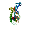
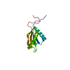
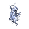
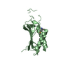
- PDB-4rlc: Crystal structure of the N-terminal beta-barrel domain of Pseudom... -

+
Open data


ID or keywords:

Loading...

-
Basic information

Entry
Database: PDB / ID: 4rlc
TitleCrystal structure of the N-terminal beta-barrel domain of Pseudomonas aeruginosa OprF
ComponentsOuter membrane porin F
KeywordsTRANSPORT PROTEIN / outer membrane protein / beta-barrel
Function / homology
Function and homology information


adhesion of symbiont to host / complement component C3b binding / outer membrane / porin activity / pore complex / monoatomic ion transport / cell outer membrane / regulation of cell shape / calcium ion binding
Similarity search - Function
Outer membrane porin F, N-terminal / OprF membrane domain / TSP type-3 repeat / Porin - #20 / Outer membrane protein, OmpA-like, conserved site / OmpA-like domain. / Outer membrane protein, bacterial / OmpA-like domain superfamily / OmpA family / OmpA-like domain ...Outer membrane porin F, N-terminal / OprF membrane domain / TSP type-3 repeat / Porin - #20 / Outer membrane protein, OmpA-like, conserved site / OmpA-like domain. / Outer membrane protein, bacterial / OmpA-like domain superfamily / OmpA family / OmpA-like domain / OmpA-like domain profile. / Outer membrane protein/outer membrane enzyme PagP, beta-barrel / Porin / Beta Barrel / Mainly Beta
Similarity search - Domain/homology
Outer membrane porin F
Similarity search - Component
Biological speciesPseudomonas aeruginosa (bacteria)
MethodX-RAY DIFFRACTION / SYNCHROTRON / MOLECULAR REPLACEMENT / Resolution: 1.6 Å
AuthorsZahn, M. / Basle, A. / van den Berg, B.
CitationJournal: J.Mol.Biol. / Year: 2015
Title: Small-Molecule Transport by CarO, an Abundant Eight-Stranded beta-Barrel Outer Membrane Protein from Acinetobacter baumannii.
Authors: Zahn, M. / D'Agostino, T. / Eren, E. / Basle, A. / Ceccarelli, M. / van den Berg, B.
History
DepositionOct 16, 2014Deposition site: RCSB / Processing site: RCSB
Revision 1.0Apr 22, 2015Provider: repository / Type: Initial release
Revision 1.1Jun 24, 2015Group: Database references
Revision 1.2Feb 28, 2024Group: Data collection / Database references / Derived calculations
Category: chem_comp_atom / chem_comp_bond ...chem_comp_atom / chem_comp_bond / database_2 / struct_ref_seq_dif / struct_site
Item: _database_2.pdbx_DOI / _database_2.pdbx_database_accession ..._database_2.pdbx_DOI / _database_2.pdbx_database_accession / _struct_ref_seq_dif.details / _struct_site.pdbx_auth_asym_id / _struct_site.pdbx_auth_comp_id / _struct_site.pdbx_auth_seq_id

-
Structure visualization

Structure viewerMolecule:
MolmilJmol/JSmol

Downloads & links

-
Assembly

Deposited unit
A: Outer membrane porin F
hetero molecules


Theoretical massNumber of molelcules
Total (without water)21,6998
Polymers19,5541
Non-polymers2,1457
Water52229
1


  • Idetical with deposited unit
  • defined by author
TypeNameSymmetry operationNumber
identity operation1_555x,y,z1
Unit cell
Length a, b, c (Å)52.009, 26.779, 54.892
Angle α, β, γ (deg.)90.00, 95.79, 90.00
Int Tables number3
Space group name H-MP121

-
Components

#1: Protein Outer membrane porin F


Mass: 19553.502 Da / Num. of mol.: 1 / Fragment: N-terminal domain (UNP residues 25-184)
Source method: isolated from a genetically manipulated source
Source: (gene. exp.) Pseudomonas aeruginosa (bacteria) / Gene: oprF, PA1777 / Production host: Escherichia coli (E. coli) / References: UniProt: P13794
#2: Chemical
ChemComp-C8E / (HYDROXYETHYLOXY)TRI(ETHYLOXY)OCTANE


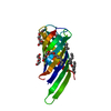
Mass: 306.438 Da / Num. of mol.: 7 / Source method: obtained synthetically / Formula: C16H34O5 / Comment: C8E, detergent*YM
#3: Water ChemComp-HOH / water


Mass: 18.015 Da / Num. of mol.: 29 / Source method: isolated from a natural source / Formula: H2O

-
Experimental details

-
Experiment

ExperimentMethod: X-RAY DIFFRACTION / Number of used crystals: 1

-
Sample preparation

CrystalDensity Matthews: 1.94 Å3/Da / Density % sol: 36.76 %
Crystal growTemperature: 292 K / Method: vapor diffusion, sitting drop / pH: 4
Details: 33% PEG200, 0.2 M ammonium sulfate, 0.02 M sodium chloride, 0.02 M sodium citrate, pH 4.0, VAPOR DIFFUSION, SITTING DROP, temperature 292K

-
Data collection

DiffractionMean temperature: 100 K
Diffraction sourceSource: SYNCHROTRON / Site: Diamond / Beamline: I03 / Wavelength: 0.97957 Å
DetectorType: DECTRIS PILATUS 6M / Detector: PIXEL / Date: Dec 7, 2013
RadiationMonochromator: double crystal Si(111) / Protocol: SINGLE WAVELENGTH / Monochromatic (M) / Laue (L): M / Scattering type: x-ray
Radiation wavelengthWavelength: 0.97957 Å / Relative weight: 1
ReflectionResolution: 1.6→54.612 Å / Num. obs: 20209 / % possible obs: 99.6 %
Reflection shellResolution: 1.6→1.63 Å / % possible all: 99.4

-
Processing

Software
NameVersionClassification
GDAdata collection
MOLREPphasing
REFMAC5.8.0073refinement
XDSdata reduction
XDS/ XSCALEdata scaling
RefinementMethod to determine structure: MOLECULAR REPLACEMENT / Resolution: 1.6→54.61 Å / Cor.coef. Fo:Fc: 0.961 / Cor.coef. Fo:Fc free: 0.935 / SU B: 5.231 / SU ML: 0.08 / Cross valid method: THROUGHOUT / ESU R: 0.087 / ESU R Free: 0.092 / Stereochemistry target values: MAXIMUM LIKELIHOOD / Details: HYDROGENS HAVE BEEN ADDED IN THE RIDING POSITIONS
RfactorNum. reflection% reflectionSelection details
Rfree0.22879 1022 5.1 %RANDOM
Rwork0.18853 ---
obs0.19065 19182 99.39 %-
Solvent computationIon probe radii: 0.8 Å / Shrinkage radii: 0.8 Å / VDW probe radii: 1.2 Å / Solvent model: MASK
Displacement parametersBiso mean: 30.718 Å2
Baniso -1Baniso -2Baniso -3
1--2.12 Å20 Å2-0.42 Å2
2--4.6 Å20 Å2
3----2.35 Å2
Refinement stepCycle: LAST / Resolution: 1.6→54.61 Å
ProteinNucleic acidLigandSolventTotal
Num. atoms1045 0 87 29 1161
Refine LS restraints
Refine-IDTypeDev idealDev ideal targetNumber
X-RAY DIFFRACTIONr_bond_refined_d0.020.0191157
X-RAY DIFFRACTIONr_bond_other_d0.0010.021107
X-RAY DIFFRACTIONr_angle_refined_deg2.041.9841526
X-RAY DIFFRACTIONr_angle_other_deg0.80232544
X-RAY DIFFRACTIONr_dihedral_angle_1_deg7.2475133
X-RAY DIFFRACTIONr_dihedral_angle_2_deg37.63724.64356
X-RAY DIFFRACTIONr_dihedral_angle_3_deg13.35815160
X-RAY DIFFRACTIONr_dihedral_angle_4_deg11.827153
X-RAY DIFFRACTIONr_chiral_restr0.130.2148
X-RAY DIFFRACTIONr_gen_planes_refined0.010.021275
X-RAY DIFFRACTIONr_gen_planes_other0.0010.02280
X-RAY DIFFRACTIONr_nbd_refined
X-RAY DIFFRACTIONr_nbd_other
X-RAY DIFFRACTIONr_nbtor_refined
X-RAY DIFFRACTIONr_nbtor_other
X-RAY DIFFRACTIONr_xyhbond_nbd_refined
X-RAY DIFFRACTIONr_xyhbond_nbd_other
X-RAY DIFFRACTIONr_metal_ion_refined
X-RAY DIFFRACTIONr_metal_ion_other
X-RAY DIFFRACTIONr_symmetry_vdw_refined
X-RAY DIFFRACTIONr_symmetry_vdw_other
X-RAY DIFFRACTIONr_symmetry_hbond_refined
X-RAY DIFFRACTIONr_symmetry_hbond_other
X-RAY DIFFRACTIONr_symmetry_metal_ion_refined
X-RAY DIFFRACTIONr_symmetry_metal_ion_other
X-RAY DIFFRACTIONr_mcbond_it1.9291.611541
X-RAY DIFFRACTIONr_mcbond_other1.9271.611540
X-RAY DIFFRACTIONr_mcangle_it2.8392.374671
X-RAY DIFFRACTIONr_mcangle_other2.8382.375672
X-RAY DIFFRACTIONr_scbond_it4.0212.662616
X-RAY DIFFRACTIONr_scbond_other4.0182.664617
X-RAY DIFFRACTIONr_scangle_it
X-RAY DIFFRACTIONr_scangle_other6.1843.61856
X-RAY DIFFRACTIONr_long_range_B_refined8.13515.4681114
X-RAY DIFFRACTIONr_long_range_B_other8.13315.491115
X-RAY DIFFRACTIONr_rigid_bond_restr
X-RAY DIFFRACTIONr_sphericity_free
X-RAY DIFFRACTIONr_sphericity_bonded
LS refinement shellResolution: 1.6→1.642 Å / Total num. of bins used: 20
RfactorNum. reflection% reflection
Rfree0.302 81 -
Rwork0.301 1371 -
obs--99.11 %
Refinement TLS params.

Method: refined / Refine-ID: X-RAY DIFFRACTION

IDL112)L122)L132)L222)L232)L332)S11 (Å °)S12 (Å °)S13 (Å °)S21 (Å °)S22 (Å °)S23 (Å °)S31 (Å °)S32 (Å °)S33 (Å °)T112)T122)T132)T222)T232)T332)Origin x (Å)Origin y (Å)Origin z (Å)
11.4408-0.0565-0.61680.3329-0.15050.9470.0138-0.0228-0.0815-0.0368-0.02740.0461-0.0095-0.13870.01350.09540.01010.0010.0484-0.01170.141510.80782.383527.1627
27.0276-1.2071-2.01020.20740.35758.14630.06040.5701-0.1616-0.0092-0.09810.02810.0866-0.18490.03770.0185-0.00390.0070.0576-0.02380.015211.67642.589310.0426
31.83340.1167-0.21670.258-0.24580.2422-0.02450.1115-0.1917-0.01520.04530.02990.0272-0.0499-0.02080.0775-0.0078-0.00630.0146-0.01460.09987.408-1.883326.6947
42.721-0.8022-4.42051.3021.19377.21740.0799-0.10910.1041-0.01290.0821-0.1533-0.10180.1405-0.16210.0424-0.015-0.00530.1606-0.00530.01955.10352.6862-1.8498
52.4155-0.101-0.05370.3693-0.12440.04630.0032-0.0025-0.2218-0.0007-0.0272-0.0736-0.00790.00780.0240.09590.00380.01320.0104-0.00660.131611.0905-3.402824.5189
Refinement TLS group
IDRefine-IDRefine TLS-IDAuth asym-IDAuth seq-ID
1X-RAY DIFFRACTION1A1 - 45
2X-RAY DIFFRACTION2A46 - 68
3X-RAY DIFFRACTION3A69 - 99
4X-RAY DIFFRACTION4A100 - 109
5X-RAY DIFFRACTION5A110 - 160

+
About Yorodumi

-
News

-
Feb 9, 2022. New format data for meta-information of EMDB entries

New format data for meta-information of EMDB entries

  • Version 3 of the EMDB header file is now the official format.
  • The previous official version 1.9 will be removed from the archive.

Related info.:EMDB header

External links:wwPDB to switch to version 3 of the EMDB data model

-
Aug 12, 2020. Covid-19 info

Covid-19 info

URL: https://pdbjlc1.pdbj.org/emnavi/covid19.php

New page: Covid-19 featured information page in EM Navigator.

Related info.:Covid-19 info / Mar 5, 2020. Novel coronavirus structure data

+
Mar 5, 2020. Novel coronavirus structure data

Novel coronavirus structure data

Related info.:Yorodumi Speices / Aug 12, 2020. Covid-19 info

External links:COVID-19 featured content - PDBj / Molecule of the Month (242):Coronavirus Proteases

+
Jan 31, 2019. EMDB accession codes are about to change! (news from PDBe EMDB page)

EMDB accession codes are about to change! (news from PDBe EMDB page)

  • The allocation of 4 digits for EMDB accession codes will soon come to an end. Whilst these codes will remain in use, new EMDB accession codes will include an additional digit and will expand incrementally as the available range of codes is exhausted. The current 4-digit format prefixed with “EMD-” (i.e. EMD-XXXX) will advance to a 5-digit format (i.e. EMD-XXXXX), and so on. It is currently estimated that the 4-digit codes will be depleted around Spring 2019, at which point the 5-digit format will come into force.
  • The EM Navigator/Yorodumi systems omit the EMD- prefix.

Related info.:Q: What is EMD? / ID/Accession-code notation in Yorodumi/EM Navigator

External links:EMDB Accession Codes are Changing Soon! / Contact to PDBj

+
Jul 12, 2017. Major update of PDB

Major update of PDB

  • wwPDB released updated PDB data conforming to the new PDBx/mmCIF dictionary.
  • This is a major update changing the version number from 4 to 5, and with Remediation, in which all the entries are updated.
  • In this update, many items about electron microscopy experimental information are reorganized (e.g. em_software).
  • Now, EM Navigator and Yorodumi are based on the updated data.

External links:wwPDB Remediation / Enriched Model Files Conforming to OneDep Data Standards Now Available in the PDB FTP Archive

-
Yorodumi

Thousand views of thousand structures

  • Yorodumi is a browser for structure data from EMDB, PDB, SASBDB, etc.
  • This page is also the successor to EM Navigator detail page, and also detail information page/front-end page for Omokage search.
  • The word "yorodu" (or yorozu) is an old Japanese word meaning "ten thousand". "mi" (miru) is to see.

Related info.:EMDB / PDB / SASBDB / Comparison of 3 databanks / Yorodumi Search / Aug 31, 2016. New EM Navigator & Yorodumi / Yorodumi Papers / Jmol/JSmol / Function and homology information / Changes in new EM Navigator and Yorodumi

Read more