SEQUENCE THE CONSTRUCT WAS EXPRESSED WITH A PURIFICATION TAG MGSDKIHHHHHHENLYFQG. THE TAG WAS ... SEQUENCE THE CONSTRUCT WAS EXPRESSED WITH A PURIFICATION TAG MGSDKIHHHHHHENLYFQG. THE TAG WAS REMOVED WITH TEV PROTEASE LEAVING ONLY A GLYCINE (0) FOLLOWED BY THE TARGET SEQUENCE.
Resolution: 2.1→27.308 Å / Num. obs: 13007 / % possible obs: 99.3 % / Observed criterion σ(I): -3 / Biso Wilson estimate: 51.74 Å2 / Rmerge(I) obs: 0.038 / Net I/σ(I): 15.82
Reflection shell
Resolution (Å)
Rmerge(I) obs
Mean I/σ(I) obs
Num. measured obs
Num. unique obs
Diffraction-ID
% possible all
2.1-2.17
0.717
2
8271
2199
1
98.2
2.17-2.26
0.508
2.9
9601
2500
1
99.8
2.26-2.36
0.396
3.7
9048
2356
1
99.8
2.36-2.49
0.311
4.6
9632
2507
1
99.9
2.49-2.64
0.221
6.7
8893
2317
1
100
2.64-2.85
0.135
10.3
9426
2462
1
100
2.85-3.13
0.074
17
9047
2354
1
99.9
3.13-3.59
0.038
27.1
9296
2423
1
99.7
3.59-4.51
0.024
39
9004
2362
1
99.3
4.51-27.308
0.022
45.2
8880
2361
1
96.1
-
Phasing
Phasing
Method: MAD
-
Processing
Software
Name
Version
Classification
NB
REFMAC
5.2.0019
refinement
PHENIX
refinement
SHELX
phasing
MolProbity
3beta29
modelbuilding
XSCALE
datascaling
PDB_EXTRACT
3
dataextraction
MAR345
CCD
datacollection
XDS
datareduction
SHELXD
phasing
SHARP
phasing
Refinement
Method to determine structure: MAD / Resolution: 2.1→27.308 Å / Cor.coef. Fo:Fc: 0.961 / Cor.coef. Fo:Fc free: 0.943 / SU B: 9.222 / SU ML: 0.121 / TLS residual ADP flag: LIKELY RESIDUAL / Cross valid method: THROUGHOUT / σ(F): 0 / ESU R: 0.167 / ESU R Free: 0.168 Stereochemistry target values: MAXIMUM LIKELIHOOD WITH PHASES Details: 1. HYDROGENS HAVE BEEN ADDED IN THE RIDING POSITIONS. 2. ATOM RECORD CONTAINS RESIDUAL B FACTORS ONLY. 3. A MET-INHIBITION PROTOCOL WAS USED FOR SELENOMETHIONINE INCORPORATION DURING PROTEIN ...Details: 1. HYDROGENS HAVE BEEN ADDED IN THE RIDING POSITIONS. 2. ATOM RECORD CONTAINS RESIDUAL B FACTORS ONLY. 3. A MET-INHIBITION PROTOCOL WAS USED FOR SELENOMETHIONINE INCORPORATION DURING PROTEIN EXPRESSION. THE OCCUPANCY OF THE SE ATOMS IN THE MSE RESIDUES WAS REDUCED TO 0.75 TO ACCOUNT FOR THE REDUCED SCATTERING POWER DUE TO PARTIAL S-MET INCORPORATION. 4. RESIDUES 0 TO 2, 103 TO 115, 137 TO 145, 171 TO 193 ARE DISORDERED AND NOT MODELED IN THE STRUCTURE.
Rfactor
Num. reflection
% reflection
Selection details
Rfree
0.259
631
4.9 %
RANDOM
Rwork
0.204
-
-
-
obs
0.207
12970
99.46 %
-
Solvent computation
Ion probe radii: 0.8 Å / Shrinkage radii: 0.8 Å / VDW probe radii: 1.2 Å / Solvent model: MASK
Displacement parameters
Biso mean: 56.22 Å2
Baniso -1
Baniso -2
Baniso -3
1-
-0.05 Å2
0 Å2
0 Å2
2-
-
-0.05 Å2
0 Å2
3-
-
-
0.09 Å2
Refinement step
Cycle: LAST / Resolution: 2.1→27.308 Å
Protein
Nucleic acid
Ligand
Solvent
Total
Num. atoms
1076
0
0
42
1118
Refine LS restraints
Refine-ID
Type
Dev ideal
Dev ideal target
Number
X-RAY DIFFRACTION
r_bond_refined_d
0.017
0.022
1120
X-RAY DIFFRACTION
r_bond_other_d
0.001
0.02
755
X-RAY DIFFRACTION
r_angle_refined_deg
1.682
1.986
1519
X-RAY DIFFRACTION
r_angle_other_deg
0.925
3
1856
X-RAY DIFFRACTION
r_dihedral_angle_1_deg
4.527
5
150
X-RAY DIFFRACTION
r_dihedral_angle_2_deg
31.126
24.419
43
X-RAY DIFFRACTION
r_dihedral_angle_3_deg
13.745
15
189
X-RAY DIFFRACTION
r_dihedral_angle_4_deg
20.623
15
9
X-RAY DIFFRACTION
r_chiral_restr
0.093
0.2
178
X-RAY DIFFRACTION
r_gen_planes_refined
0.005
0.02
1262
X-RAY DIFFRACTION
r_gen_planes_other
0.001
0.02
202
X-RAY DIFFRACTION
r_nbd_refined
0.208
0.3
179
X-RAY DIFFRACTION
r_nbd_other
0.201
0.3
724
X-RAY DIFFRACTION
r_nbtor_refined
0.168
0.5
525
X-RAY DIFFRACTION
r_nbtor_other
0.094
0.5
603
X-RAY DIFFRACTION
r_xyhbond_nbd_refined
0.142
0.5
60
X-RAY DIFFRACTION
r_symmetry_vdw_refined
0.179
0.3
14
X-RAY DIFFRACTION
r_symmetry_vdw_other
0.359
0.3
29
X-RAY DIFFRACTION
r_symmetry_hbond_refined
0.295
0.5
8
X-RAY DIFFRACTION
r_mcbond_it
2.166
3
769
X-RAY DIFFRACTION
r_mcbond_other
0.489
3
303
X-RAY DIFFRACTION
r_mcangle_it
3.499
5
1195
X-RAY DIFFRACTION
r_scbond_it
6.29
8
388
X-RAY DIFFRACTION
r_scangle_it
8.888
11
324
LS refinement shell
Resolution: 2.1→2.154 Å / Total num. of bins used: 20
Rfactor
Num. reflection
% reflection
Rfree
0.403
53
-
Rwork
0.293
882
-
obs
-
935
98.32 %
Refinement TLS params.
Method: refined / Origin x: 54.534 Å / Origin y: 23.346 Å / Origin z: 10.278 Å
11
12
13
21
22
23
31
32
33
T
-0.3326 Å2
-0.0415 Å2
-0.0417 Å2
-
-0.2806 Å2
0.0336 Å2
-
-
-0.1998 Å2
L
3.319 °2
2.6145 °2
-1.1479 °2
-
4.8026 °2
-1.924 °2
-
-
2.3208 °2
S
-0.1504 Å °
0.3481 Å °
0.3173 Å °
-0.2503 Å °
0.1718 Å °
0.2502 Å °
-0.138 Å °
0.0642 Å °
-0.0214 Å °
+
About Yorodumi
-
News
-
Feb 9, 2022. New format data for meta-information of EMDB entries
New format data for meta-information of EMDB entries
Version 3 of the EMDB header file is now the official format.
The previous official version 1.9 will be removed from the archive.
In the structure databanks used in Yorodumi, some data are registered as the other names, "COVID-19 virus" and "2019-nCoV". Here are the details of the virus and the list of structure data.
Jan 31, 2019. EMDB accession codes are about to change! (news from PDBe EMDB page)
EMDB accession codes are about to change! (news from PDBe EMDB page)
The allocation of 4 digits for EMDB accession codes will soon come to an end. Whilst these codes will remain in use, new EMDB accession codes will include an additional digit and will expand incrementally as the available range of codes is exhausted. The current 4-digit format prefixed with “EMD-” (i.e. EMD-XXXX) will advance to a 5-digit format (i.e. EMD-XXXXX), and so on. It is currently estimated that the 4-digit codes will be depleted around Spring 2019, at which point the 5-digit format will come into force.
The EM Navigator/Yorodumi systems omit the EMD- prefix.
Related info.:Q: What is EMD? / ID/Accession-code notation in Yorodumi/EM Navigator
Yorodumi is a browser for structure data from EMDB, PDB, SASBDB, etc.
This page is also the successor to EM Navigator detail page, and also detail information page/front-end page for Omokage search.
The word "yorodu" (or yorozu) is an old Japanese word meaning "ten thousand". "mi" (miru) is to see.
Related info.:EMDB / PDB / SASBDB / Comparison of 3 databanks / Yorodumi Search / Aug 31, 2016. New EM Navigator & Yorodumi / Yorodumi Papers / Jmol/JSmol / Function and homology information / Changes in new EM Navigator and Yorodumi
 Movie
Movie Controller

 Yorodumi
 Open data
 Basic information
 Components
 Keywords
 Function and homology information
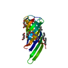
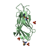
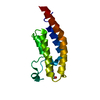
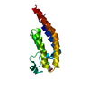
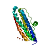
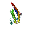
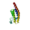
 Silicibacter pomeroyi DSS-3 (bacteria)
 X-RAY DIFFRACTION /
  Authors
 Citation
 Structure visualization
 Downloads & links
 Other downloads


 PDBj
PDBj Assembly
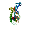

 Mass: 18.015 Da / Num. of mol.: 42 / Source method: isolated from a natural source / Formula: H2O
 Sample preparation
 / Beamline: BL9-2 / Wavelength: 0.91162, 0.97922, 0.97905
 Processing