[English] 日本語
 Yorodumi
- PDB-4rjy: Crystal structure of E. coli L-Threonine Aldolase in complex with... -
+ Open data
- Basic information
| Entry | Database: PDB / ID: 4rjy | ||||||
|---|---|---|---|---|---|---|---|
| Title | Crystal structure of E. coli L-Threonine Aldolase in complex with a non-covalently uncleaved bound L-serine substrate | ||||||
 Components Components | Low specificity L-threonine aldolase | ||||||
 Keywords Keywords | LYASE / Pyridoxal-5-phosphate / threonine aldolase / aldimine / catalytic mechanism / retro-aldol cleavage / PLP-dependent enzymes | ||||||
| Function / homology | Aspartate Aminotransferase, domain 1 / Aspartate Aminotransferase, domain 1 / Aspartate Aminotransferase; domain 2 / Type I PLP-dependent aspartate aminotransferase-like (Major domain) / Alpha-Beta Complex / 3-Layer(aba) Sandwich / Alpha Beta / SERINE / :  Function and homology information Function and homology information | ||||||
| Biological species |  | ||||||
| Method |  X-RAY DIFFRACTION / X-RAY DIFFRACTION /  MOLECULAR REPLACEMENT / Resolution: 2.1 Å MOLECULAR REPLACEMENT / Resolution: 2.1 Å | ||||||
 Authors Authors | Safo, M.K. / Chowdhury, N. / Gandhi, A.K. | ||||||
 Citation Citation |  Journal: Biochim.Biophys.Acta / Year: 2015 Journal: Biochim.Biophys.Acta / Year: 2015Title: Molecular basis of E. colil-threonine aldolase catalytic inactivation at low pH. Authors: Remesh, S.G. / Ghatge, M.S. / Ahmed, M.H. / Musayev, F.N. / Gandhi, A. / Chowdhury, N. / di Salvo, M.L. / Kellogg, G.E. / Contestabile, R. / Schirch, V. / Safo, M.K. | ||||||
| History |
|
- Structure visualization
| Structure viewer | Molecule:  Molmil Molmil Jmol/JSmol Jmol/JSmol |
|---|
- Downloads & links
- Download
| PDBx/mmCIF format |  4rjy.cif.gz 4rjy.cif.gz | 277.6 KB | Display |  PDBx/mmCIF format PDBx/mmCIF format |
|---|---|---|---|---|
| PDB format |  pdb4rjy.ent.gz pdb4rjy.ent.gz | 232.8 KB | Display |  PDB format PDB format |
| PDBx/mmJSON format |  4rjy.json.gz 4rjy.json.gz | Tree view |  PDBx/mmJSON format PDBx/mmJSON format | |
| Others |  Other downloads Other downloads |
-Validation report
| Summary document |  4rjy_validation.pdf.gz 4rjy_validation.pdf.gz | 474.9 KB | Display |  wwPDB validaton report wwPDB validaton report |
|---|---|---|---|---|
| Full document |  4rjy_full_validation.pdf.gz 4rjy_full_validation.pdf.gz | 522.1 KB | Display | |
| Data in XML |  4rjy_validation.xml.gz 4rjy_validation.xml.gz | 64.3 KB | Display | |
| Data in CIF |  4rjy_validation.cif.gz 4rjy_validation.cif.gz | 88 KB | Display | |
| Arichive directory |  https://data.pdbj.org/pub/pdb/validation_reports/rj/4rjy https://data.pdbj.org/pub/pdb/validation_reports/rj/4rjy ftp://data.pdbj.org/pub/pdb/validation_reports/rj/4rjy ftp://data.pdbj.org/pub/pdb/validation_reports/rj/4rjy | HTTPS FTP |
-Related structure data
| Similar structure data |
|---|
- Links
Links
- Assembly
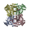
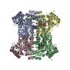
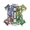
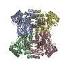
| Deposited unit | 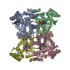
| ||||||||
|---|---|---|---|---|---|---|---|---|---|
| 1 |
| ||||||||
| Unit cell |
|
- Components
| #1: Protein | Mass: 36756.883 Da / Num. of mol.: 4 Source method: isolated from a genetically manipulated source Source: (gene. exp.)   #2: Chemical | ChemComp-NA / #3: Chemical | ChemComp-SER / #4: Water | ChemComp-HOH / | |
|---|
-Experimental details
-Experiment
| Experiment | Method:  X-RAY DIFFRACTION / Number of used crystals: 1 X-RAY DIFFRACTION / Number of used crystals: 1 |
|---|
- Sample preparation
| Crystal | Density Matthews: 2.33 Å3/Da / Density % sol: 47.31 % |
|---|---|
| Crystal grow | Temperature: 298 K / Method: vapor diffusion, hanging drop / pH: 5.6 Details: Freshly dialyzed eTA (22 mg/mL in 20 mM potassium phosphate, pH 7.0) was incubated with L-serine (6.25 mM), precipitant solution contains 0.1 M sodium citrate tribasic dihydrate, pH 5.6, 20% ...Details: Freshly dialyzed eTA (22 mg/mL in 20 mM potassium phosphate, pH 7.0) was incubated with L-serine (6.25 mM), precipitant solution contains 0.1 M sodium citrate tribasic dihydrate, pH 5.6, 20% v/v 2-propoanol, 20% v/v PEG 4000, VAPOR DIFFUSION, HANGING DROP, temperature 298K |
-Data collection
| Diffraction | Mean temperature: 100 K | ||||||||||||||||||||||||||||||||||||||||||||||||||||||||||||||||||||||||||||||||||||||||
|---|---|---|---|---|---|---|---|---|---|---|---|---|---|---|---|---|---|---|---|---|---|---|---|---|---|---|---|---|---|---|---|---|---|---|---|---|---|---|---|---|---|---|---|---|---|---|---|---|---|---|---|---|---|---|---|---|---|---|---|---|---|---|---|---|---|---|---|---|---|---|---|---|---|---|---|---|---|---|---|---|---|---|---|---|---|---|---|---|---|
| Diffraction source | Source:  ROTATING ANODE / Type: RIGAKU MICROMAX-007 / Wavelength: 1.5417 Å ROTATING ANODE / Type: RIGAKU MICROMAX-007 / Wavelength: 1.5417 Å | ||||||||||||||||||||||||||||||||||||||||||||||||||||||||||||||||||||||||||||||||||||||||
| Detector | Type: RIGAKU RAXIS IV++ / Detector: IMAGE PLATE / Date: Nov 22, 2011 | ||||||||||||||||||||||||||||||||||||||||||||||||||||||||||||||||||||||||||||||||||||||||
| Radiation | Monochromator: Ni Filter / Protocol: SINGLE WAVELENGTH / Monochromatic (M) / Laue (L): M / Scattering type: x-ray | ||||||||||||||||||||||||||||||||||||||||||||||||||||||||||||||||||||||||||||||||||||||||
| Radiation wavelength | Wavelength: 1.5417 Å / Relative weight: 1 | ||||||||||||||||||||||||||||||||||||||||||||||||||||||||||||||||||||||||||||||||||||||||
| Reflection | Resolution: 2.1→32.8 Å / Num. obs: 77165 / % possible obs: 98 % / Observed criterion σ(F): 2 / Observed criterion σ(I): 2 / Redundancy: 2.91 % / Rmerge(I) obs: 0.093 / Χ2: 0.87 / Net I/σ(I): 8.4 / Scaling rejects: 24943 | ||||||||||||||||||||||||||||||||||||||||||||||||||||||||||||||||||||||||||||||||||||||||
| Reflection shell | Diffraction-ID: 1
|
- Processing
| Software |
| ||||||||||||||||||||||||||||
|---|---|---|---|---|---|---|---|---|---|---|---|---|---|---|---|---|---|---|---|---|---|---|---|---|---|---|---|---|---|
| Refinement | Method to determine structure:  MOLECULAR REPLACEMENT / Resolution: 2.1→30 Å / σ(F): 0 / Stereochemistry target values: Engh & Huber MOLECULAR REPLACEMENT / Resolution: 2.1→30 Å / σ(F): 0 / Stereochemistry target values: Engh & Huber
| ||||||||||||||||||||||||||||
| Solvent computation | Bsol: 78.7595 Å2 | ||||||||||||||||||||||||||||
| Displacement parameters | Biso max: 113.59 Å2 / Biso mean: 27.2876 Å2 / Biso min: 1.07 Å2
| ||||||||||||||||||||||||||||
| Refinement step | Cycle: LAST / Resolution: 2.1→30 Å
| ||||||||||||||||||||||||||||
| Refine LS restraints |
| ||||||||||||||||||||||||||||
| Xplor file |
|
 Movie
Movie Controller





 PDBj
PDBj


